6go6ckt5b8|3000F7576AC3|Tab_Articles|Fulltext|0xf1ffc825010000006700000001000700
6go6ckt5b5idvals|123
6go6ckt5b5idcol1|ID
6go6ckt5b5|2000F757Tab_Articles|Fulltext
A 21 day old female child presented in pediatric OPD with complaints of multiple limb deformities noted since birth. Antenatal period was uneventful and the two antenatal ultrasounds done at 18 weeks and 32 weeks failed to show any abnormality. Child was delivered vaginally and post natal period remained uneventful. On examination, multiple constrictive rings in right upper limb leading to finger deformities were noted (Fig. 1). It was also associated with club foot deformities in bilateral lower limbs. A clinical diagnosis of Streeter’s dysplasia was made.
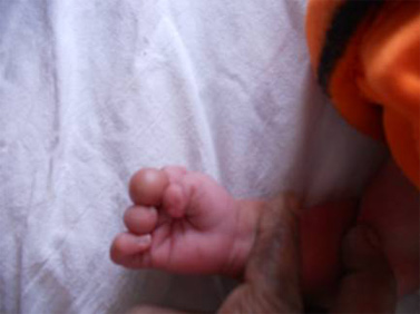
Fig 1. Streeter’s Dysplasia: Constrictive annular deformities in right hand
Streeter’s dysplasia, also known as amniotic band syndrome is characterized by multiple annular ring constriction deformities. It is a rare abnormality that manifests at birth in form of constriction rings predominantly involving distal parts of upper and lower limbs. The deformities vary from simple constriction rings to bony and soft tissue fusion along with lymphedema. Some deformities are extremely severe to lead to intrauterine amputations.[1]
Children born with Streeter’s dysplasia are usually full term or a few weeks premature; in most cases, the pregnancies are uncomplicated. Club foot is noted in nearly 25% cases, mostly resistant in nature leading to ambulatory difficulties.[2] Other associated findings include encephalocele, cleft lip or palate, renal abnormalities, cardiac defects, hemihypertrophy, anterolateral bowing of the tibia and tibial pseudoarthrosis.[3] Detailed investigations like X-ray limbs, ultrasound abdomen and ECHO did not reveal any associated malformation except for club foot.
The most accepted hypothesis about Streeter’s dysplasia states that incomplete obliteration of the extra coelomic space renders the amnion fragile and subject to spontaneous or traumatic rupture. The floating amniotic band encircles body parts leading to constriction deformities. Also, extravasation of amniotic fluid leads to transient oligohydramnios allowing developing fetus to have very little space to move. This may contribute to the severity of clubfeet deformities seen with amniotic band syndrome.[4]
Indications for intervention depend on the medical stability of the child and on the neurovascular status of the limb. Of all the deformities, only the tight constriction bands with gross lymphedema, vascular compromise, or both necessitate immediate surgical release. Clubfeet should be manipulated and early casts should be put with regular follow up. Surgical intervention should be initiated as in any idiopathic club foot.
References
-
K. Kawamura, K. Chung. Constriction Band Syndrome
Hand Clinics 2009; 25(2): 257-264
-
Hennigan, SP, Kuo, KN. Resistant talipes equinovarus associated with congenital constriction band syndrome.
J Pediatr Orthop. 2000; 20(2): 240-245
-
Tanguy AF, Dalens BJ, Boisgard S. Congenital constricting band with pseudoarthrosis of the tibia and fibula. A case report.
J Bone Joint Surg 1995; 77A(8): 1251-1254.
-
Ross MG. Pathogenesis of amniotic band syndrome.
Am J Obstet Gynecol. Aug 2007; 197(2): 219-220
If you like forever classic fake watches, you cannot miss the uk top quality cheap replica rolex watches canada.
UK Omega Replica Watches For Men