Introduction
Lipomas are the most common benign mass of mesenchymal origin [
1]. It consists of mature adipocytes, often encapsulated by a thin fibrous tissue and typically lies in the subcutaneous plane [
2]. Only 13% of them arises in the head and neck region, most commonly in the posterior neck [
1]. Lipoma in the temporal region is rare with two documented cases on sieving through the literature [
3]. In this region, the lipoma may arise from the layers of temporal fat, the anatomy of which has been discussed further. This case report aims to contribute valuable insights and challenges into the anatomy, management, and outcomes of lipomas occurring in the temporal region.
Case Report
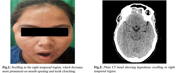
A 29-year-old female presented with a complaint of swelling in the right temporal region for the past one year [Fig.1]. The swelling was not associated with any pain, erythema or discharge. Her primary concern regarding the swelling was cosmesis. No complaint of similar swelling elsewhere in the body was elicited. She had no other known co-morbidities. On examination, a fullness was noted in the right anterior temporal region. On clenching teeth, a prominent well defined 3 × 3 cm firm, non-tender swelling appeared above the zygoma. The overlying skin was normal and no pulsations were felt over the swelling. The NCCT head revealed a well-defined thick-walled round hypodense lesion measuring 26 × 22 mm in the right temporal region with calcific foci within [Fig.2]. Underlying bone appeared normal. Fine needle aspiration cytology showed fragments of mature adipose tissue in hemorrhagic background suggestive of lipoma.
She underwent excision of the lesion under local anaesthesia through a remote cosmetic hairline incision and the tissue was sent for histopathological examination [Fig.3,4]. The lipoma was found deep to the superficial temporal fascia during the surgery. The frontal branch of facial nerve was identified and preserved. Histopathological examination of the excised tissue confirmed lipoma. At the follow-up visit, the swelling was seen to be completely resolved [Fig.5,6].
Discussion
The layers of tissue in the temporal region consists of skin, subcutaneous tissue, superficial temporal fascia, loose areolar tissue, deep temporal fascia, temporalis muscle, pericranium and cranium. This loose areolar tissue, also known as innominate fascia is a relatively avascular plane which facilitates movement of superficial scalp over deeper muscular fascial layers [
4]. There are three pads of fat interspersed between these layers of fascia. Fat lying deep to the superficial layer of fascia is superficial layer of fat, between the two layers of deep temporalis fascia is intermediate layer of fat and beneath the deep layer of fascia is deep layer of fat [
5]. Lipomas can arise from any of these three layers. In our reported case, it was seen to be arising from the intermediate temporal fat pad. Intramuscular lipomas arising from the temporalis muscle have been reported as well [
6].
In addition to contributing to the literature, this article draws attention to the anatomy of temporal region as this is important in variety of surgeries involving temporo-mandibular joint and malar arch, harvesting of temporoparietal fascia flap, pterional craniotomy, face-lift surgeries etc. Frontal branch of facial nerve crosses this region and damage to this branch can cause temporal hollowing and facial asymmetry. Hence, meticulous dissection in the desired plane along with knowledge of surgical anatomy of the frontal branch should be known to the operating surgeon. Pitenguy’s line is an imaginary line drawn 0.5 cm below the tragus to 1.5 cm lateral and above the lateral eyebrow. This line marks the surface anatomy of frontal/temporal branch of facial nerve, which lies in the SMAS plane [
4].
Lipoma in this region can be accessed through a cosmetic hairline incision. Al-Kayat and Bramley approached this region through a pre-auricular incision which gives a good accessibility as well as protects the facial branch [7]. Remote scar placement results in more favourable cosmetic outcome by eliminating perceptible scar. Techniques such as “pot-lid” technique and liposuction has been used to decrease the incidence of cosmetic disfigurement post removal of these masses [
8]. Although CT scan can also detect lipomas, MRI is considered more accurate for evaluation of extent, localisation and characterization of lipomatous lesion in uncommon sites [
9]. Recurrence rate of lipoma is considered to be low, if excised completely.
Conclusion
By presenting a rare clinical case, we intend to shed light on the challenges faced by surgeons when dealing with such atypical presentations of lipoma in specific anatomical locations, such as the temporal region.
Contributors: AKS: manuscript editing, patient management; RRN: manuscript writing, patient management; RRN will act as a study guarantor. Both authors approved the final version of this manuscript and are responsible for all aspects of this study.
Funding: None; Competing interests: None stated.
References
- Som PM, Scherl MP, Rao VM, Biller HF. Rare presentations of ordinary lipomas of the head and neck: a review. Am J Neuroradiol. 1986;7(4):657-664.
- Charifa A, Azmat CE, Badri T. Lipoma Pathology. In: StatPearls. Treasure Island (FL): StatPearls Publishing; 2022. Available from: http://www.ncbi.nlm.nih.gov/books/NBK482343/
- Davies J, Srinivasan B, Brennan PA. Lipoma of the temporal region: a rare case series. Ann R Coll Surg Engl. 2021;103(1):e42-43.
- Green Sanderson K, Conti A, Colussi M, Connolly C. A simple clinical application for locating the frontotemporal branch of the facial nerve using the zygomatic arch and the tragus. Aesthet Surg J. 2020;40(5):NP223-227.
- Bohr C, Bajaj J, Soriano RM, Shermetaro C. Anatomy, Head and Neck, Temporoparietal Fascia. In: StatPearls. Treasure Island (FL): StatPearls Publishing; 2022. Available from: http://www.ncbi.nlm.nih.gov/books/NBK507912/
- Uemura T, Suse T, Yokoyama T, Mitsukawa N, Yoshikawa A. Intramuscular benign lipoma of the temporalis muscle. Scand J Plast Reconstr Surg Hand Surg. 2002;36(4):231-234.
- Al-Kayat A, Bramley P. A modified pre-auricular approach to the temporomandibular joint and malar arch. Br J Oral Surg. 1979;17(2):91-103.
- Cillo JE, Caloss R, Wendelken JA. Excision of subcutaneous facial cysts and lipomas using cosmetic approaches. J Oral Maxillofac Surg. 2006;64(11):1603-1616.
- Cappabianca S, Colella G, Pezzullo MG, Russo A, Iaselli F, Brunese L, et al. Lipomatous lesions of the head and neck region: imaging findings in comparison with histological type. Radiol Med (Torino). 2008;113(5):758-770.