|
|
|
|
|
Giant Malignant Phylloides
|
|
|
|
Neha Baporikar, Siddhartha Verma Department of General Surgery, Rabindra Nath Tagore Medical College, Udaipur, Rajasthan, India. |
|
|
|
|
|
Corresponding Author:
|
|
Dr. Neha Baporikar Email: nehabaporikar@gmail.com |
|
|
|
|
|
|
|
|
Received:
25-MAY-2023 |
Accepted:
03-OCT-2023 |
Published Online:
05-NOV-2023 |
|
|
|
|
|
|
|
Abstract
|
|
|
|
Background: Phyllodes tumors of the breast, a rare fibroepithelial neoplasm, pose diagnostic and therapeutic challenges due to their diverse morphological spectrum and classification into benign, intermediate, and malignant variants. This abstract explores the clinical manifestations, histological features, and management complexities associated with these tumors, emphasizing the diagnostic difficulties in differentiating them from other fibroepithelial lesions. Case Report: A 35-year-old female with a Malignant Giant Phyllodes tumor, sought medical attention due to a rapidly enlarging lump in her right breast. The tumor measured 20×20×15 cm, emphasizing the diagnostic challenges posed by such substantial masses. Diagnostic procedures included fine-needle aspiration cytology (FNAC) and tru-cut biopsy, revealing the difficulties in achieving diagnostic consistency among pathologists. The patient underwent a right mastectomy, and the post-operative course was uneventful. However, the biopsy report indicated malignant Phyllodes with positive margins. Despite recommendations for revision surgery, the patient declined, highlighting the complexities in patient management. Conclusion: This case contributes valuable insights to the medical literature, emphasizing the intricate diagnostic process and surgical decisions involved in managing Malignant Giant Phyllodes tumors. |
|
|
|
|
|
Keywords :
|
Breast, Malignant, Mastectomy, Surgery, Tumor.
|
|
|
|
|
|
|
|
|
|
|
|
Introduction
Phyllodes tumor of the breast is a rare fibroepithelial neoplasm, comprising 0.3-1% of all tumors [ 1]. Its classification into benign, borderline, and malignant variants by the World Health Organization (WHO) relies on histological features, encompassing stromal cellularity, nuclear atypia, mitotic activity, and the appearance of overgrowth at tumor margins [ 1]. This rare tumor type, while typically non-metastatic, can exhibit rapid growth, reaching considerable sizes and occasionally ulcerating the skin. Typically well-demarcated, Phyllodes tumors exhibit a morphologic spectrum comprising connective tissue mixed with gelatinous cystic and solid areas [ 2]. In the hematogenous spread, Phyllodes tumors differ from other breast tumors, showing a propensity for recurrence in the local area. Hematogenous metastases, when they occur, typically involve the lung, bone, abdominal viscera, and mediastinum. Giant Phyllodes tumors, exceeding 10 cm and found in only 20% of cases, present a surgical challenge due to the potential for significant defects after complete negative margin excision [ 3]. While complete excision with negative margins is often curative, axillary dissection is usually unnecessary as these tumors do not spread via lymphatics. Diagnosing Phyllodes tumors remains challenging, particularly in distinguishing them from other fibroepithelial lesions, such as giant Fibroadenoma, both through imaging and in fine-needle aspiration cytology (FNAC) and tru-cut biopsy.
Case Report
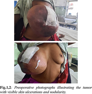
A 35-year-old woman sought medical attention for rapidly enlarging lump in her right breast over the past 6 months. Two weeks prior to seeking medical advice, the mass had ulcerated, accompanied by serosanguinous discharge and pain [Fig.1,2]. Upon admission, the patient was alert, conscious, and in stable condition. Breast examination revealed a massive ulcerating mass, approximately 20×20×15 cm in size, occupying the entire right breast. Nodularity and ulceration with sero-sanguinous and purulent discharge were evident, while no other masses or axillary lymph nodes were palpable.
The patient's hemoglobin level was 10g/dL, and other blood investigations yielded results within normal limits. Ultrasonography (USG) revealed a substantial mass involving the entire right breast with internal vascularity, along with a few sub-centimetric lymph nodes in the bilateral axillary region. Mammography depicted a homogenous dense opacity occupying the entire right breast with skin involvement. No calcifications were observed within the lesion (BIRADS III/IV). Fine-needle aspiration cytology (FNAC) was performed twice, suggesting a Phylloides tumor with mucoid degeneration but without specifying benign or malignant characteristics. Tru-cut biopsy was unsuccessful. A right mastectomy was performed to excise the entire mass, followed by primary closure [Fig.3,4]. The post-operative course was uneventful, and the patient was discharged after five days. A follow-up appointment after seven days revealed a biopsy report indicating malignant Phylloides with positive margins. Despite recommendations for revision surgery, the patient declined and was subsequently referred to Oncology for further management after suture removal.
Discussion
Phyllodes tumors, a rare class of tumors featuring mixed connective tissue and epithelial elements, present a diagnostic and surgical challenge. They are categorized as benign, intermediate, and malignant, with difficulties in cytologically differentiating low-grade benign Phyllodes from typical Fibroadenomas. These tumors exhibit a proliferation of connective tissue and ductal elements, and the malignant variant displays increased cellularity, invasive margins, and a genuinely sarcomatous appearance. Characterized as firm, lobulated masses, Phyllodes tumors range from 2 to 40 cm, with those surpassing 10 cm termed Giant Phyllodes [ 4]. Histologically, Phyllodes tumors display distinctive whorls of stroma forming large clefts lined by epithelium, resembling leaf-like structures. Coined as "cystosarcoma phyllodes," the term captures the leaf-like stromal projections into cystic spaces and the fleshy consistency of the tumor [5-8]. The stroma, more cellular than fibroadenomas, reveals bland fibroblastic cells with infrequent mitoses [6]. Recurrences involve both epithelial and connective tissue elements, while metastases predominantly comprise the malignant connective tissue component [7]. Mammographically, Phyllodes tumors resemble fibroadenomas, presenting as round densities with smooth borders and a radiolucent halo due to tissue compression. Ultrasonography (USG) reveals discrete lesions with cystic spaces [9]. Clinical diagnosis relies on large sizes, a history of rapid growth, and occurrence in older females.In the case being reported, a rapidly enlarging breast lump emphasizes the diagnostic challenges associated with these rare and substantial masses. This unique case contributes to the existing medical literature by highlighting the complexities in the clinical manifestations, diagnostic procedures, and management strategies for Malignant Giant Phyllodes tumors. Management depends on tumor classification, with benign tumors curatively treated via local excision. Intermediate or borderline tumors necessitate excision with 1 cm margins, and close follow-up is essential due to the risk of local recurrence. Despite efforts to enhance diagnostic precision, challenges persist, particularly in distinguishing Phyllodes from other fibroepithelial lesions. Fine-needle aspiration cytology (FNAC) often falls short, emphasizing the need for tru-cut biopsy, which, despite being more effective, still poses challenges in diagnostic consistency among pathologists [1]. In cases of malignant Phyllodes, en bloc surgical excision, such as total mastectomy, is recommended. Unlike many other sarcomas, regional lymph node dissection is unnecessary due to hematogenous spread. Positive margins in benign cases may warrant observation, but malignant Phyllodes cases should strongly consider revision surgery. The presented case, where the patient declined revision surgery and was subsequently referred to Oncology, emphasizes the challenges in achieving optimal management strategies.
Conclusion
The reported case highlights the diagnostic intricacies and surgical challenges posed by these rare breast masses. Achieving a disease-free state necessitates precise diagnostic approaches, with tru-cut biopsy proving more pertinent than FNAC. Despite advancements, challenges in diagnostic consistency among pathologists persist. The unique clinical presentation and management decisions in this case contribute valuable insights to the existing medical literature, emphasizing the importance of patient consent and compliance in achieving optimal outcomes.
Contributors: NB: manuscript writing, patient management; SV: manuscript editing, patient management. SV will act as a study guarantor. Both authors approved the final version of this manuscript and are responsible for all aspects of this study. Funding: None; Competing interests: None stated.
References - Zhang Y, Kleer CG. Phyllodes tumor of the breast: histopathologic features, differential diagnosis, and molecular/genetic updates. Archives of Pathology & Laboratory Medicine. 2016;140(7):665-671.
- Albalawi IA. A huge phyllodes tumor in the breast: a case report. Electronic Physician. 2018;10(6):6951.
- Fernández-Ferreira R, Arroyave-Ramírez A, Motola-Kuba D, Alvarado-Luna G, Mackinney-Novelo I, Segura-Rivera R. Giant benign mammary phyllodes tumor: report of a case and review of the literature. Case Reports in Oncology. 2021;14(1):123-133.
- Chaudhary D, Singh V, Mallya V, Mandal S, Khurana N, Singh R. Utility of trucut biopsy in diagnosing phyllodes tumor. Journal of Mid-life Health. 2019;10(3):135.
- Parker SJ, Harries S. Phyllodes tumours. Postgraduate Medical Journal. 2001;77(909):428-435.
- Yan Z, Gudi M, Lim SH. A large benign phyllodes tumour of the breast: a case report and literature review. International Journal of Surgery Case Reports. 2017;39:192-195.
- World Health Organization. Histological typing of breast tumours.Neoplasma. 1983;30(1):113-123.
- Strode M, Khoury T, Mangieri C, Takabe K. Update on the diagnosis and management of malignant phyllodes tumors of the breast. The Breast. 2017;33:91-96.
- Mishra SP, Tiwary SK, Mishra M, Khanna AK. Phyllodes tumor of breast: a review article. ISRN Surg. 2013;361469.
|
|
|
|
|
|
|
Search Google Scholar for
|
|
|
Article Statistics |
|
Baporikar N, Verma SGiant Malignant Phylloides.JCR 2023;13:110-113 |
|
Baporikar N, Verma SGiant Malignant Phylloides.JCR [serial online] 2023[cited 2026 Mar 14];13:110-113. Available from: http://www.casereports.in/articles/13/4/Giant-Malignant-Phylloides.html |

|
|
|
|
|