|
|
|
|
|
Adult Onset Epidermal Verrucous Nevus and Angiokeratoma: An Unusual Coexistence.
|
|
|
symbicort inhaler coupon
symbicort inhaler price go
Rajesh Sinha, Pallavi Sharma, Hari Shankar Pandey1
From the Department of Dermatology and Pathology1,
Govt. Medical College, Haldwani, Nanital, Uttarakhand, India.
Official Swiss Replica Watches UK replicaswiss.co.uk
|
|
|
|
|
|
Corresponding Author:
|
Dr. Rajesh Sinha
Email: sinhaderma@gmail.com
Mobile: 09627010430 |
|
|
|
|
|
|
|
|
Received:
30-MAR-2012 |
Accepted:
29-APR-2012 |
Published Online:
30-APR-2012 |
|
|
|
|
|
|
|
Abstract
|
|
|
|
Verrucous epidermal nevi are characterized by localized or diffuse, brown or gray-brown verrucous papules which may coalesce to form well-demarcated papillomatous plaques. Various lesions like squamous cell carcinoma, basal cell carcinoma, may arise on epidermal nevus but association with vascular lesion is rare except in epidermal nevus syndromes and proteus syndrome. We report a case of adult onset epidermal verrucous nevus associated with clinical and histopathological features of angiokeratoma.
2024 New Best Fake Cartier Watches: cartiershops.com
|
|
|
|
|
|
Keywords :
|
Epidermal verrucous nevus, Angiokeratoma
|
|
|
|
|
|
|
|
|
|
|
|
6go6ckt5b8|3000F7576AC3|Tab_Articles|Fulltext|0xf1ff0445010000001500000001001900
6go6ckt5b5idvals|126
6go6ckt5b5idcol1|ID
6go6ckt5b5|2000F757Tab_Articles|Fulltext
Epidermal verrucous nevus is a skin hamartoma characterized by a muliple verrucous papules coalesceing simuntaneously to form a serpiginous plaque. We report an association of adult onset epidermal verrucous nevus with angiokeratoma. Association of epidermal verrucous nevus and angiokeratoma is rare and to best of our knowledge only one case has been reported so far.[1]
Case Report
A 58 year-old man presented to outpatient department with an plaque on the right half of posterior aspect of neck. The lesion which started as a small papule, had been present for last 7 years. The lesion has slowly progressed in elevation and extent but remained localised to the back of the neck. The lesion was neither painful nor pruritic. Examination revealed an slightly elevated, hyperpigmented erythematous verrucous plaque measuring 2.5 × 7 cm, interspersed with dark red papular eruptions on right half of posterior aspect of neck, extending from hair line to the neck [Figure 1]. A clinical diagnosis of adult onset verrucous nevus was made, and skin biopsy was performed. Biopsy revealed hyperkeratosis, acanthosis, papillomatosis, and dilated capillaries in widened dermal papilliae [Figure 2].
All best models of uk wholesale Swiss cheap replica watches uk on google can be find here.
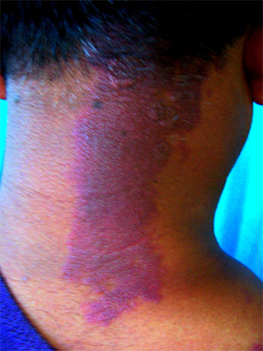 Fig.1: Verrucous erythematous plaque over posterior aspect of neck.
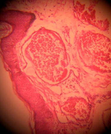 Fig.2: Histopathology of the lesion showing hyperkeratosis, acanthosis, and dilated vessels in widened dermal papillae (40X, H&E).
Discussion
Epidermal nevi are hamartomous proliferations of the epithelium and adnexal structures. Epidermal nevi may be classified into a number of variants, based on clinical morphology, extent of involvement, and the predominant epidermal structure in the lesion. Variants include verrucous epidermal nevus (keratinocytes), nevus sebaceous (sebaceous gland), nevus comedonicus (pilosebaceous unit), eccrine nevus (eccrine gland), or apocrine nevus (apocrine gland). [2,3]
Verrucous epidermal nevi are characterized by localized or diffuse, close set skin coloured brown or gray-brown verrucous papules which may coalesce to form well-demarcated papillomatous plaques. Histopathology of verrucous epidermal nevus shows hyperkeratosis, acanthosis, papilliomatosis, and elongation of rete ridges. Various lesions like squamous cell carcinoma, basal cell carcinoma, may arise on epidermal nevus but association with vascular lesion is very rare except in epidermal nevus syndromes and proteus syndrome.[4,5]
Angiokeratoma is a vascular dermatosis with a histology of subepidermal dilated blood vessels, characterized by telangiectasia and hyperkeratosis. Angiokeratoma presents as elevated, warty, dark red to purple, slightly compressible papules with hyperkeratotic scale over the surface. The lesions are pigmented due to hemosiderin pigment deposition in the dermis.The laboratory evaluation that confirms the diagnosis is a biopsy. Overall, altered hemodynamics (typically caused by trauma) appear to produce telangiectatic vessels of the papillary dermis with an overlying reactive hyperkeratosis to the epidermis.[6]
Association of epidermal verrucous nevus and angiokeratoma is very rare and pubmed search showed that only one case has been reported so far.[1] Khatami et al reported occurrence of epidermal nevus and angiokeratoma together in the form of two oval plaques over right upper thigh in a 17 year old girl.[1] Clinical manifestation of our patient showed association of verrucous epidermal nevus and angiokeratoma which was confirmed by histopathological examination. Clinical diagnosis of verrucous nevus was done on the basis of lesion morphology and mode of onset while angiokeratoma was diagnosed on basis of dark red papules. Histologically, hyperkeratosis, acanthosis, papillamatosis, are features of verrucous nevus and dialated capillaries in widened dermal papillae are features of angiokeratoma. Our case is also unique because of late onset at an age of 51 years. The case is being reported because of rarity of this association.
Fig.2: Histopathology of the lesion showing hyperkeratosis, acanthosis, and dilated vessels in widened dermal papillae (40X, H&E).
Discussion
Epidermal nevi are hamartomous proliferations of the epithelium and adnexal structures. Epidermal nevi may be classified into a number of variants, based on clinical morphology, extent of involvement, and the predominant epidermal structure in the lesion. Variants include verrucous epidermal nevus (keratinocytes), nevus sebaceous (sebaceous gland), nevus comedonicus (pilosebaceous unit), eccrine nevus (eccrine gland), or apocrine nevus (apocrine gland). [2,3]
Verrucous epidermal nevi are characterized by localized or diffuse, close set skin coloured brown or gray-brown verrucous papules which may coalesce to form well-demarcated papillomatous plaques. Histopathology of verrucous epidermal nevus shows hyperkeratosis, acanthosis, papilliomatosis, and elongation of rete ridges. Various lesions like squamous cell carcinoma, basal cell carcinoma, may arise on epidermal nevus but association with vascular lesion is very rare except in epidermal nevus syndromes and proteus syndrome.[4,5]
Angiokeratoma is a vascular dermatosis with a histology of subepidermal dilated blood vessels, characterized by telangiectasia and hyperkeratosis. Angiokeratoma presents as elevated, warty, dark red to purple, slightly compressible papules with hyperkeratotic scale over the surface. The lesions are pigmented due to hemosiderin pigment deposition in the dermis.The laboratory evaluation that confirms the diagnosis is a biopsy. Overall, altered hemodynamics (typically caused by trauma) appear to produce telangiectatic vessels of the papillary dermis with an overlying reactive hyperkeratosis to the epidermis.[6]
Association of epidermal verrucous nevus and angiokeratoma is very rare and pubmed search showed that only one case has been reported so far.[1] Khatami et al reported occurrence of epidermal nevus and angiokeratoma together in the form of two oval plaques over right upper thigh in a 17 year old girl.[1] Clinical manifestation of our patient showed association of verrucous epidermal nevus and angiokeratoma which was confirmed by histopathological examination. Clinical diagnosis of verrucous nevus was done on the basis of lesion morphology and mode of onset while angiokeratoma was diagnosed on basis of dark red papules. Histologically, hyperkeratosis, acanthosis, papillamatosis, are features of verrucous nevus and dialated capillaries in widened dermal papillae are features of angiokeratoma. Our case is also unique because of late onset at an age of 51 years. The case is being reported because of rarity of this association.
References
- Khatami A, Ghorbani Z, Gorouhi F, Davari P, Firooz A. Angiokeratoma and epidermal nevus. Eur J Dermatol. 2008; 18: 100-101.
- Thomas VD, Swanson NA, Lee KK. Benign epithelial tumours, Hamartomas, and Hyperplasias. In: Wolff K, Goldsmith LA, Katz SI, Gilchrest BA, Paller AS, Leffell DJ, editors. Fitzpatcrik's Dermatology in General Medicine.7th ed. NewYork: McGraw-Hill; 2008. pp.1054-56.
- Solomon LM, Esterly NB. Epidermal and other congenital organoid nevi. Curr Probl Pediatr 1975; 6: 1-56.
- Twede JV, Turner JT, Biesecker LG, Darling TN. Evolution of skin lesions in Proteus syndrome. J Am Acad Dermatol 2005; 52: 834-838.
- Calzavara Pinton P, Carlino A, Manganoni AM, Donzelli C, Facchetti F. Epidermal nevus syndrome with multiple vascular hamartomas and malformations. G Ital Dermatol Venereol 1990; 125: 251-254.
- Schiller PI, Itin PH. Angiokeratomas: an update. Dermatology. 1996; 193: 275-282.
|
|
|
|
|
|
|
Search Google Scholar for
|
|
|
Article Statistics |
|
Sinha R, Sharma P, Pandey HSAdult Onset Epidermal Verrucous Nevus and Angiokeratoma: An Unusual Coexistence..JCR 2012;2:6-8 |
|
Sinha R, Sharma P, Pandey HSAdult Onset Epidermal Verrucous Nevus and Angiokeratoma: An Unusual Coexistence..JCR [serial online] 2012[cited 2026 Mar 31];2:6-8. Available from: http://www.casereports.in/articles/2/1/adult-onset-epidermal-verrucous.html |

|
|
|
|
|