|
|
|
|
|
Congenital Hyporhinia with Associated Malformations
|
|
|
viagra diskuze viagra prodej inzerce Ngamdu YB, Ibrahim BM1, Bello M2, C Tahir, Kodiya AM, Ashiru MG2, Sandabe MB,Garandawa HI, Isa A.
Department of ENT Surgery; Burns and Plastic unit,
Department of Surgery1; Department of Paediatrics2
University of Maiduguri Teaching Hospital, Maiduguri. |
|
|
|
|
|
Corresponding Author:
|
Dr. YB Ngamdu
Email: ybugam@yahoo.com |
|
|
|
|
|
|
|
|
Received:
29-JUL-2012 |
Accepted:
10-AUG-2012 |
Published Online:
05-SEP-2012 |
|
|
|
|
|
|
|
Abstract
|
|
|
|
Congenital hyporhinia, also known as partial arhinia, is a very rare congenital abnormality of nasal embryogenesis with unknown aetiology. It is commonly associated with other craniofacial anomalies. A one day old neonate presented to our facility with hypoplastic nasal pyramid, hypertelorism, microcephaly, microphthalmia, micrognathia and cleft palate. A case of congenital hyporhinia with associated anomalies in tropics is presented. |
|
|
|
|
|
Keywords :
|
Congential hyporhinia, Arhinia, Neonate
|
|
|
|
|
|
|
|
|
|
|
|
6go6ckt5b8|3000F7576AC3|Tab_Articles|Fulltext|0xf1ff94aa01000000b500000001000900 6go6ckt5b5idvals|142 6go6ckt5b5idcol1|ID 6go6ckt5b5|2000F757Tab_Articles|Fulltext Congenital hyporhinia (partial arhinia) is an extremely uncommon congenital nasal anomaly. About 40 cases of the congenital arhinia that have been reported in the English literature since 1931, 4 of which were congenital hyporhinia [1,2]. The cause is still unknown, most cases are sporadic but also few familial cases have been reported [3]. This anomaly is usually found to be associated with other malformations involving the craniofacial area, palate clefts, ear defects, central nervous system. Problems with feeding, severe airway and phonetics usually accompany congenital hyporhinia in paediatric age [1]. A case of congenital hyporhinia associated with microcephaly, microphthalmia, cleft palate, micrognathia and hyperthelorism is reported.
Case Report
Attention of otorhinolaryngologist was drawn to review a day old full term female neonate, with nasal malformation delivered vaginally to 34 year old para 5 mother. Pregnancy was unsupervised and baby was delivered at home under the care of traditional birth attendant. Parents were not consanguineously married. Mother had no significant history of ingestion of any traditional or orthodox medicines or exposure to radiation during her pregnancy, and remained well throughout. She does not smoke or drink alcohol and has never abused drugs.
Examination revealed birth weight of 2.8 kg, birth length of 49 cm and head circumference of 29 cm (microcephaly). External nasal pyramid was hypoplastic and collapsed, with remnants of alar cartilage barely palpable and a centrally placed single stenotic anterior nasal nare. Columella, nasal septum and the philtrium were absent [Figure 1]. Insertion of nasogastric tube revealed anterior nare leading to 5 mm deep single nasal cavity with atretic posterior choanae [Figure 2]. Hypertelorism, microphthalmia, micrognathia and cleft palate were noted. Echocardiography did not reveal any abnormality.
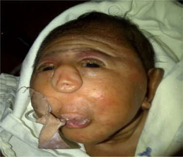 Figure 1 showing hypertelorism, collapsed and flabby nasal pyramid, single centrally placed stenotic anterior nasal nare with an oxygen catheter in the oral cavity.
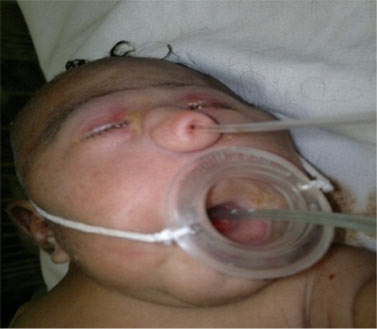 Figure 2 showing size 8G nasogastric tube in single anterior nasal opening and the improvised Mac Govern nipple for feeding and breathing
An improvised Mac Govern nipple was put in place for feeding and breathing [Figure 2].Parents could not afford further investigations like computerized tomography and chromosomal analysis to delineate extent and cause of malformation. After detailed and compassionate discussion with parents, patient was discharged after 6 days of admission but defaulted follow up after the first visit.
Discussion
Congenital hyporhinia is a rare defect of embryogenesis often associated with other congenital anomalies. Hyporhinia and associated congenital anomalies often have a significant effect on the immediate and long-term outcome of the neonate. Prenatal diagnostic advances have greatly improved the possibility of early detection of congenital anomalies. This gives not only an opportunity to plan and improve perinatal care but also offers the option to terminate the pregnancy for cases in which the prognosis is likely to be poor. This was not possible in our case as pregnancy was unsupervised.
Figure 2 showing size 8G nasogastric tube in single anterior nasal opening and the improvised Mac Govern nipple for feeding and breathing
An improvised Mac Govern nipple was put in place for feeding and breathing [Figure 2].Parents could not afford further investigations like computerized tomography and chromosomal analysis to delineate extent and cause of malformation. After detailed and compassionate discussion with parents, patient was discharged after 6 days of admission but defaulted follow up after the first visit.
Discussion
Congenital hyporhinia is a rare defect of embryogenesis often associated with other congenital anomalies. Hyporhinia and associated congenital anomalies often have a significant effect on the immediate and long-term outcome of the neonate. Prenatal diagnostic advances have greatly improved the possibility of early detection of congenital anomalies. This gives not only an opportunity to plan and improve perinatal care but also offers the option to terminate the pregnancy for cases in which the prognosis is likely to be poor. This was not possible in our case as pregnancy was unsupervised.
The primitive face develops from five facial prominences. One of these, the frontonasal prominence, is responsible for nasal development during the third to tenth weeks of gestation [4]. Joseph et al in 2003 reported a new classification system dedicated solely to congenital nasal anomalies [5]. It takes its origins from Whitaker’s classification scheme for craniofacial deformities and is similarly a simple morphogenic classification system based on aetiology, anatomy and treatment principles. All congenital nasal anomalies were classified into:
Type I- hypoplasia and atrophy
Type II- Hyperplasia and duplication
Type III - Clefts
Type IV – Neoplasm and vascular anomalies
Type I anomalies were found with an incidence of 62%, this represent paucity, atrophy, or underdevelopment of skin, subcutaneous tissue, muscle, cartilage, and/or bone [6]. The present report falls into type I Whitaker’s classification scheme.
The aetiology of arhinia is unknown and attempts have been made to associate it with maternal diabetes, hypertension, and toxaemia of pregnancy [7]. In present case the mother was neither diabetic nor hypertensive. The pregnancy was uneventful as in other reported cases [8]. Although most cases are sporadic, few familial cases have been reported. Mostly the karyotype is normal. Genetic aberrations of chromosome 9 and chromosome 3- 12 have been detected in few cases [3,9,10]. Others believe arhinia results from failed Invagination of nasal placodes, leading to impaired nasal cavity formation [5].
The cases may have associated anomalies in the central nervous system, craniofacial area, other midline defect, extremities and spine. These associated congenital anomalies determine the immediate and long term outcome [11]. Radiological evaluation is the gold standard in the evaluation and proper surgical planning[11]. In the present report, no radiological evaluation was carried out due to financial constraint.
There is controversy about the correct timing and the surgical procedure but the surgery is staged. In the early days of life endotracheal intubation or tracheostomy had been done to relief upper airway obstruction. Surgical reconstruction should be delayed until preschool age in most cases [12]. In our case, no surgical intervention was necessary. In this index case is difficult to comment either because patient defaulted follow up after the first visit. Long term outcomes of these cases have not been reported.
When life threating congenital malformations is detected, a detailed discussion with the parents is essential. It is difficult time for parents, and they will require social support and counselling, including a risk assessment for future pregnancies. Several ethical and economic issues arise in determining life and death issues. The issue of the child's best interest is probably the most important. Is surgery for deformities ethical when it will not change the ultimate prognosis? The answer to the question is probably not, since the surgery is not likely to improve the child's quality of life.
References
- MacGlone L. Congenital arhinia. J Paediatr. Child Health.2003; 39: 474-476.
- Robert MG, Manuel GV, Adolfo BC, Elina EM. Congenital partial arhinia with no associated malformations. 2009;4:162-164.
- Hou JW. Congenital arhinia with de novo reciprocal translocation, t(3;12) (q13,2;p11. 2). Am J Med Genet. 2004;130A:2000-2003.
- Farag MM, Ragaeie A, El-Oteify MA. Rare midline congenital anomalies of the face. J. Laryngol. Otol. 1988;102:1040.
- Joseph EL, Richard EK, Linton AW, Scott PB. Congenital nasal anomalies: A classification scheme. 2000;113: 676-689.
- de Blécourt RA, Roddi R, Berg JP, Bloem JJ. Nasal dysplasia. Ann Plast Surg. 1996;37:633-637.
- Kemble J. The importance of the nasal septum in facial development. J Laryngol Otol. 1973;87:379.
- Cusick W, Sullivan CA, Rojas B, Poole AE, Poole DA. Prenatal diagnosis of total arhinia. Ultrasound Obstet. Gynecol 2000;15:259-261.
- Kaminker CP, Dain L, Lamas MA, Sanchez JM. Brief clinical report: Mosaic trisomy 9 syndrome with unsual phenotype. Am J. Med Genet. 1985;22:237-241.
- Cohen D, Goitein K. Arhinia revisited. Rhinology 1987;25:237-244.
- Olsen E, Gjelland K, Reigstad H, Rosendahi K. Congenital absence of the nose: A case report and literature review. Pediatr. Radiol 2001;31:225-232.
- Mathur NN, Dubey NK, Kumar S, Bothra R. Chadha A. Arhinia. Int. J. Pediat Otorhinol. 2005;69:97-99.
|
|
|
|
|
|
|
Search Google Scholar for
|
|
|
Article Statistics |
|
Ngamdu YB, Ibrahim BM, Bello M, C Tahir, Kodiya AM, Ashiru MG, Sandabe MB,Garandawa HI, Isa A.Congenital Hyporhinia with Associated Malformations.JCR 2012;2:54-56 |
|
Ngamdu YB, Ibrahim BM, Bello M, C Tahir, Kodiya AM, Ashiru MG, Sandabe MB,Garandawa HI, Isa A.Congenital Hyporhinia with Associated Malformations.JCR [serial online] 2012[cited 2026 May 21];2:54-56. Available from: http://www.casereports.in/articles/2/2/congenital-hyporhinia-with.html |

|
|
|
|
|