|
|
|
|
|
Perineal Ectopic Testis with Unilateral Scrotal Hypoplasia
|
|
|
Adittya K. Sharma, Fareed Rehan, RP Sanjay, CS Ratkal
From the Department of Urology, Institute of NephroUrology, Bangalore, Karnataka, India. |
|
|
|
|
|
Corresponding Author:
|
Dr. Adittya K. Sharma
Email: dradityaks@gmail.com |
|
|
|
|
|
|
|
|
Received:
9-JAN-2013 |
Accepted:
16-JAN-2013 |
Published Online:
15-FEB-2013 |
|
|
|
|
|
|
|
Abstract
|
|
|
|
The ectopic testis is frequently misdiagnosed as cryptorchidism or anorchism. We present a case of three month old male infant diagnosed as right ectopic perineal testis and right hemiscrotal hypoplasia. Ectopic testes are thought to be greater at risk of trauma, testicular torsion, subfertility and malignancy. Although definitive evidence in support of these fears in case of perineal ectopia is lacking due to its rarity and lack of long follow-up. Surgical correction of undescended testes is generally done at about 6 months of age to allow for spontaneous descent. On the other hand there is no need to delay surgery in ectopic testis because possible descent as seen in undescended testis will not occur. Yet the timing of surgery can be individualized for perineal ectopia without any unnecessary delay.
|
|
|
|
|
|
Keywords :
|
Cryptorchidism, Gonadal Dysgenesis, Scrotum, Perineum, Infertility.
|
|
|
|
|
|
|
|
|
|
|
|
6go6ckt5b8|3000F7576AC3|Tab_Articles|Fulltext|0xf1ffb47502000000b201000001000800 6go6ckt5b5idvals|180 6go6ckt5b5idcol1|ID 6go6ckt5b5|2000F757Tab_Articles|Fulltext Introduction
Cryptorchidism is the most common glandular anomaly. It is noted in 3% of male infants at birth [1]. Ectopic testis (ET) occurs in only about 5% of the cases of empty scrotum. Perineal ectopic testis, the most common type of maldescended testis occurs about 1% of the patients [2].
Case Report
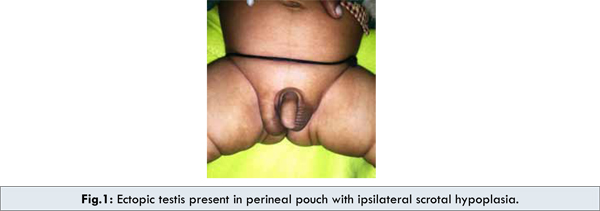
A three month old male infant presented with abnormally shaped scrotum and right testis abnormally placed in a outpouching below and lateral to expected right hemiscrotum. Baby was first child and with no familial history or consanguinity. On examination infant was having hypoplastic right hemiscrotum and normal left half of scrotum with normally palpable left testis [Fig.1]. There was firm mass identical like left testis palpable in a perineal pouch on right side. Ultrasound revealed normal left testis with right testis in perineal pouch. Hence, a diagnosis of right ectopic perineal testis with hypoplastic right hemiscrotum was made. Patient was planned for right side orchidopexy in dartos pouch later.
Discussion
The ectopic testis is frequently misdiagnosed as cryptorchidism or even anorchism. Similar pathological findings in ectopic and undescended testes as well as the association of ectopic testis with a contralateral undescended testis suggest that ectopic and undescended testes are variants of the same congenital anomaly. Thus, boys with ectopic testis may have an increased incidence of subfertility and testicular malignancy. This spectrum of abnormal testicular position and its range of pathological conditions and complications may appropriately be called the undescended testis sequence [1]. The condition is thought to result from a deviation of terminal testicular descent from its usual path with the testes becoming lodged in various abnormal locations. Five major sites of ectopia are perineum, femoral canal, superficial pouch, suprapubic area and contralateral pouch. In addition to these well recognized sites, preperitoneal, extracorporeal and the anterior abdominal wall ectopic testes have been reported [3].
Testicular descent can be described in two phases: transabdominal and inguinoscrotal. During the inguinoscrotal phase, the testis may deviate from the normal path of descent and migrate to an abnormal location; this is called ectopic testes. If the testis cannot be palpated in the usual position or in the groin near the external inguinal ring, all the probable sites for an ectopic testis should be meticulously examined.
Different hypotheses have been proposed regarding etiologies of ectopic testis. It is felt to be either the result of hormonal imbalance between androgen and calcitonin gene-related peptide (CGPR), or the result of aberrant gubernacular stabilization due to an anomaly at its distal end, or possibly be caused by local mechanical obstacles blocking the normal descent [1].
Ectopic testes are thought to be greater at risk of trauma, testicular torsion, subfertility and malignancy. But due to low incidence there isn’t enough evidence showing increased risk in long term follow-up of perineal ectopic testis. Subfertility should not be concern for unilateral perineal ectopia. Surgical correction of undescended testes is generally done at about 6 months of age to allow for spontaneous descent. If there is co-existing inguinal hernia, early intervention is needed to prevent hernia incarceration. It’s been suggested that there is no need to delay surgery in ectopic testis because possible descent as seen in undescended testis will not occur [4]. Although it is arguable as there are conflicting evidence about reduction of relative risk of malignancy, between orchidopexy done in early child hood versus any time before puberty. Further studies are needed to clarify the relationship between age at orchidopexy and risk of testicular cancer [5]. It is still unclear whether there should be similar urgency to operate in cases of perineal ectopia which already lies outside abdomen and is available for examination [6].
Conclusion
Perineal testicular ectopia is rare and correction although not as difficult as in cases of undescended testis due to long pedicle yet there is no consensus regarding exact timing of correction. In authors opinion timing can be individualized without any unnecessary delay.
References
- Hutcheson JC, Snyder HM 3rd, Zuñiga ZV, Zderic SA, Schultz DJ, Canning DA, Huff DS. Ectopic and undescended testes: 2 variants of a single congenital anomaly? J Urol. 2000;163:961-963.
- Nounla J, Tröbs RB, Rolle U. Perineal ectopic testis: a rare cause of empty scrotum. Urol Int. 2001;67:246-248.
- Rao PL, Gupta V, Kumar V. Anterior abdominal wall--an unusual site for ectopic testis. Pediatr Surg Int. 2005;21:687-688.
- Celayir AC, Sander S, Eliçevik M. Timing of surgery in perineal ectopic testes: analysis of 16 cases. Pediatr Surg Int. 2001;17:167-168.
- Barthold JS. Abnormalities of the testes and scrotum and their surgical management. In: Wein AJ (eds). Campbell-Walsh Urology.10th ed. Philadelphia, Pa: Saunders Elsevier; 2011:chap 132.
- Alonso Domínguez F, Osorio Acosta V. A rare case of testicular ectopia. Arch Esp Urol. 2004; 57:547-549.
|
|
|
|
|
|
|
Search Google Scholar for
|
|
|
Article Statistics |
|
K. Sharma A, Rehan F, Sanjay RP, Ratkal CSPerineal Ectopic Testis with Unilateral Scrotal Hypoplasia.JCR 2013;3:68-70 |
|
K. Sharma A, Rehan F, Sanjay RP, Ratkal CSPerineal Ectopic Testis with Unilateral Scrotal Hypoplasia.JCR [serial online] 2013[cited 2026 May 26];3:68-70. Available from: http://www.casereports.in/articles/3/1/perineal-ectopic-testis.html |

|
|
|
|
|