|
|
|
|
|
Modified Cantwell-Ransley Repair of Male Penopubic Epispadias: Report of Two Cases and Review of the Literature
|
|
|
|
Bijit Lodh, Somarendra Khumukcham, Bernard Amer, Kaku Akoijam Singh
From the Department of Urology, Regional Institute of Medical Sciences (RIMS), Imphal, Manipur, India. |
|
|
|
|
|
Corresponding Author:
|
|
Dr. Bijit Lodh
Email: drblodh@yahoo.co.in |
|
|
|
|
|
|
|
|
Received:
12 AUG 2013 |
Accepted:
9-SEP-2013 |
Published Online:
25-SEP-2013 |
|
|
|
|
|
|
|
Abstract
|
|
|
|
The reported incidence of male epispadias is 1 in 1,17,000 male births. The majority of cases go unseen in surgeons’ life because of uncommon existence of this anomaly. We present our experience using the modified Cantwell-Ransley technique; particularly focusing on postoperative anatomical and functional outcomes. Between 2011 and 2013, two boys having primary penopubic epispadias with the spraying of urinary stream underwent one stage surgical reconstruction. Cavernocavernostomy was performed in all cases as a part of a procedure to correct dorsal curvature and for penile lengthening, but none of them required bladder neck procedure. At 3rd and 6th month follow-up period both of them found to have significant step forward subjective and objective outcomes. The cosmetic appearance of the phallus was not satisfactory in one case and it is because post-operatively the patient developed necrotic patches over the glans and neo-urethral skin that healed with fibrosis. |
|
|
|
|
|
Keywords :
|
Epispadias, Male Genitalia, Urinary Bladder, Penis, Reconstructive Surgical Procedures, Urethra.
|
|
|
|
|
|
|
|
|
|
|
|
6go6ckt5b8|3000F7576AC3|Tab_Articles|Fulltext|0xf1fff80b040000002602000001000700 6go6ckt5b5idvals|246 6go6ckt5b5idcol1|ID 6go6ckt5b5|2000F757Tab_Articles|Fulltext Introduction
Epispadias is a rare congenital anomaly characterized by a dorsal urethral defect where it is replaced by a broad mucosal strip lining the dorsum of the penis extending toward the bladder with the potential incompetence of the sphincter mechanism. In contrary to hypospadias, the current entity is not frequently discussed in the society of urology, plastic and paediatric surgery, and it is because of its uncommon occurrence. Associated anomalies in patients with epispadias are deformed external genitalia, diastasis of the pubic symphysis and urinary incontinence. Nevertheless, a carefully constructed and well-planned approach is the prerequisite to the management of these patients. The objectives of repair must include achievement of urinary continence, cosmetically acceptable genitalia, normal genital function, and preservation of fertility potential in most cases. Here, we are interested to report such cases not only for its rarity but also to refresh our memory with respect to its surgical repair and outcome.
Case Report
Two boys, aged 5 and 9 year underwent surgical repair for correction of epispadias in the department of Urology, RIMS, over a period of eighteen months (Sept 2011 to March 2013). Pre-operatively both patients had mild stress urinary dribbling and spraying of the urine stream. Significant dorsal cordee (20 degree) was observed in one patient. There was no abnormal widening of the pubic symphysis in any of the cases. Other physical, laboratory and radiological imaging were unremarkable. A flaccid unstretched penile length was measured from the penopubic junction to the tip of the penis in the standing position. We assessed the penile length preoperatively and at 3 months postoperatively. The Wilcoxon signed-rank test was used to determine the difference in the preoperative and postoperative penile lengths. All patients were operated under general anaesthesia. The operative technique used was Modified Cantwell-Ransley technique. The procedure involves placement of traction stitches into the glans penis [Fig.1]. A reverse meatal advancement and granuloplasty (MAGPI) or Ipgam procedure performed at the distal urethral plate [Fig. 2]. After the initial urethral strip incision on the lateral edges of the urethral plate and around the epispadiac meatus the skin is dissected off and glans wings are created [Fig.3]. The urethra is tubularized using a continuous running suture (5-0 chromic) over a No. 8 Fr infant feeding tube. Corporal bodies are incised dorsally at the point of maximum curvature that provides a diamond shaped extension of the incision [Fig.4]. The corpora are then closed over the neourethra with two running suture of 5-0 polydiaxone, and the diamond-shaped defects in the adjacent area of the corpora are sutured to each other. Glans wings and the skin are re-approximated with interrupted 5-0 polyglycolic sutures/chromic catgut [Fig.5]. A Z-plasty at the base of the penis is closed with interrupted 5-0 polyglycolic sutures/chromic catgut. Oxybutynin (0.1-0.15 mg/kg per dose) is started immediately after surgery to decrease the incidence of bladder spasms and enhance the patient’s comfort. The dressing was opened on the 7th post-operative day and the catheter were removed on the 12th post-operative day. The patients underwent regular follow-up examinations at 1, 3 and 6 months and were evaluated for subjective and objective outcomes.
Operative outcome measures are shown in Table 1. Post-operatively penis appeared longer in both cases. However, no statistically significant inference on pre and postoperative unstretched penile length could be derived on such small number of patients (P=0.068). All the patients had acceptable conical shaped glans. The peno-pubic angle varied from 120-135 degree in the flaccid state in standing position [Fig. 6]. No patient developed post-operative fistula and none of them, required urethral dilatation. The cosmetic appearance of the penis was excellent in one patient and none of the patient had a problem of dorsal cordee. The results of their adequate sexual activity were not judged because of the age limiting factor. Postoperative voiding was devoid of urine spraying.
Discussion
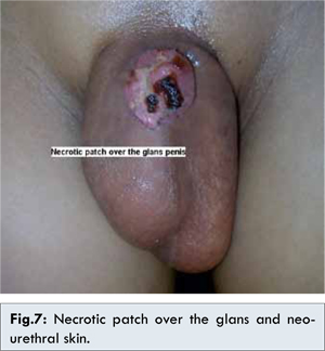
The first recorded case of epispadias is attributed to the Byzantine Emperor Heraclius for his unknown disease [ 1, 2]. The combined incidence of the Exstrophy-epispadias complex (EEC) spectrum can be estimated at 1 in 10,000 births and isolated male epispadias defect is present in 10% of cases of EEC [ 3, 4]. Although epispadias is considered to be the least severe defect of the exstrophy–epispadias complex, the treatment of this anomaly is far from trivial and repair can be challenging. There are two popular surgical techniques described well in the literature with their pros and cons; the first is the modified Cantwell technique, which involves partial disassembly of the penis and placement of the urethra in a more normal position and the second and most recent evolution is the complete penile disassembly. The drawback of modified Ransley techniques are persistence of short penile length and residual dorsal cordee that is likely to be eliminated in complete penile disassembly [ 5, 6]. The major disadvantages of the Mitchell technique for epispadias repair are the necessity for aggressive dissection and occasional resultant of hypospadias meatus that require 2nd stage urethroplasty [ 7]. This is because the urethral plate is usually shorter than the corpora cavernosa. Here, we have presented our preliminary experience with the modified Cantwell technique for isolated penopubic epispadias to determine the lessons that have been learned. On the first post operative dressing one patient was found to have necrotic patch and slough over the glans and neo-urethral skin [Fig.7]. This is the reason why the post-operative appearance was not satisfactory in one patient because healing occurred with secondary intention. Urethrocutaneous fistulas and urethral strictures represent the most common complications following the modified Cantwell-Ransley procedure. Surer and associates [ 8] evaluated their patients three months post-operatively and found that fistula and stricture rate was 19% and 10% respectively. Ransley and colleagues reported a fistula rate of 4% and urethral stricture rate of 5.3% [ 9]. Although our series was small but no patient developed urethrocutaneous fistula that is in concurrence with the findings of Pippi Salle JL et al [ 10]. In our case, neo-meatus were created at its orthotropic position, therefore voiding through an ectopic meatus was possible to avoid. Most boys (up to 70%) with penopubic epispadias, the bones of the pelvis are widely separated. This affects the bladder neck and external sphincter leading to incompetence and constant or stress urinary dribbling [ 2, 7]. There was no abnormal widening of the pubic symphysis in our case and none of the patient had urinary incontinence. Therefore, we have not considered for bladder neck reconstruction.
Conclusion
Although surprising, but it is not uncommon to evident an isolated continent penopubic epispadias. Here, we have reported such entity. Modified Cantwell Ransley repair was best suited for them and found to have satisfactory outcome.
References
- Lascaratos J, Poulakou-Rembelakou E, Rembelakos A, Marketos S. The first case of epispadias: an unknown disease of the Byzantine Emperor Heraclius (610–641 AD). British Journal of Urology 1995;76:380–383.
- Gearhart JP, Mathews RI. Exstrophy-epispadias complex. In: Wein AJ, editor. Campbell-Walsh Urology, 10th ed. Saunders Elsevier: Philadelphia; 2007. pp. 3325.
- Ebert AK, Reutter H, Ludwig M, Rosch WH. The Exstrophy-epispadias complex. Orphanet Journal of Rare Diseases. 2009;4:23.
- Mokhless I, Youssif M, Ismail HR, Higazy H. Partial penile disassembly for isolated epispadias repair. Urology. 2008;71:235-238.
- Narsimhan KL, Mohanty SK, Singh N, Samujh R, Rao KLN, Mitra SK. Epispadias repair using the Mitchell technique. J Pediatr Surg. 1999;34:461-463.
- Ransley PG, Duffy PG. Bladder exstrophy closure and epispadias repair. In: Spitz L, Coran AG, editors. Rob & Smith Operative Surgery (Pediatric Surgery). Chapman & Hall: London; 1995. pp. 745-759.
- Sarin YK, Manchanda V. Mitchell’s technique for epispadias repair: Our preliminary experience. J Indian Assoc Pediatr Surg. 2006; 11:31-34.
- Surer I, Baker LA, Jeffs RD, Gearhart JP. The modified Cantwell-Ransley repair for exstrophy and epispadias: 10year experience. J Urol. 2000;164:1040-1043.
- Gearhart JP, Mathews RI. Exstrophy-epispadias complex. In: Wein AJ, editor. Campbell-Walsh Urology, 10th ed. Saunders Elsevier: Philadelphia; 2007. pp. 3357.
- Pippi Salle JL, Jednak R, Capolicchio JP, França IMP, Labbie A, Gosalbez R. A ventral rotational skin flap to improve cosmesis and avoid chordee recurrence in epispadias repair. BJU International. 2002;90:918–923.
|
|
|
|
|
|
|
Search Google Scholar for
|
|
|
Article Statistics |
|
Lodh B, Khumukcham S, Amer B, Singh KAModified Cantwell-Ransley Repair of Male Penopubic Epispadias: Report of Two Cases and Review of the Literature.JCR 2013;3:344-348 |
|
Lodh B, Khumukcham S, Amer B, Singh KAModified Cantwell-Ransley Repair of Male Penopubic Epispadias: Report of Two Cases and Review of the Literature.JCR [serial online] 2013[cited 2026 May 2];3:344-348. Available from: http://www.casereports.in/articles/3/2/Modified-Cantwell-Ransley-Repair-of-Male-Penopubic-Epispadias-Report-of-Two-Cases-and-Review-of-the-Literature.html |

|
|
|
|
|