6go6ckt5b8|3000F7576AC3|Tab_Articles|Fulltext|0xf1ff68a6030000000902000001000700
6go6ckt5b5idvals|225
6go6ckt5b5idcol1|ID
6go6ckt5b5|2000F757Tab_Articles|Fulltext
Introduction
Porphyria cutanea tarda (PCT) is the most common subtype of porphyria [
1]. The disorder results from low levels of the enzyme uroporphyrinogen decarboxylase (UROD). The disease is characterized by blistering of the skin in areas that receive higher levels of exposure to sunlight. While a deficiency in this enzyme is the direct cause leading to this disorder, there are a number of both genetic and environmental risk factors that are associated with PCT [
2]. We report a 32 year patient with porphyria cutanea tarda who presented with multiple blisters and was diagnosed with coral red fluorescence of urine under Wood’s lamp.
Case Report
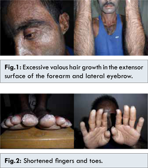
A 32 years old Asian, nondiabetic, normotensive male, seeked medical attention for frequent appearance of multiple blisters over different parts of the body with burning sensation of skin during exposure to sunlight for last 15 years. These blisters first appeared on face, gradually progressed to scalp, dorsum of hands, feet, forearm and legs. Blisters ruptured easily releasing clear fluid, healed by scarring with some hypo or hyper pigmentation and this phenomenon increased during work, summer season and hot bath and relatively decreased during winter. He also noticed excessive growth of hair [Fig.1] over the face most prominently over the eyebrows, forehead, malar area, temporal area, ear and nose along with feeling of tightness of skin of both hands and feet and shortening of all fingers and toes [Fig.2] for last 5 years. He complained of burning sensation and easy fatigibility on exposure to sunlight.
There was no history of color change of his digits on exposure to cold. Also there was no history of arthritis, oral ulceration, butterfly rash, or intake of any offending drug. With these problems the patient consulted with many doctors, kabiraj (local traditional doctors), homeopaths, village quacks and treated accordingly without any significant improvement.
His general examination revealed no abnormalities but examination findings of integumentary system showed tense blister present over his right dorsum of foot which had negative Nikolsky sign, some depigmented melasma like patch present over his face and forehead without telangiectasia or atrophy with thickness over skin of palm and soles. Excessive body hair was present on areas exposed to sun. He had deformed nails of fingers and toes, subungual hyperkeratosis of both fingers and toes and onycholysis of several toe nails.
His routine blood examination findings were unremarkable. Liver function test was normal and anti HCV serology was negative. On bed side urine examination, coral red fluorescence was seen under Wood’s lamp [Fig.3]. He was planned for skin biopsy for histopathology and serum porphyrin level with 24 hours urine for porphyrin level estimation. But as these tests were not available in Chittagong and the poor patient was unable to do it from distant laboratory, the patient was diagnosed as a case of Porphyria cutanea tarda on clinical grounds.
Discussion
Patients who are ultimately diagnosed with PCT first seek treatment following the development of photosensitivity in the form of blisters and erosions on commonly exposed areas of the skin. This is usually observed in the face, hands, forearms, and lower legs. It heals slowly with scarring. Though blisters are the most common skin manifestations of PCT, other skin manifestations like hyperpigmentation (as if they are getting a tan) and hypertrichosis (mainly on top of the cheeks) also occur. PCT is a chronic condition, with external symptoms often subsiding and recurring as a result of a number of factors [
3]. In the 20% of cases, porphyria cutanea tarda is inherited as an autosomal dominant pattern [
3].
High levels of uroporphyrinogen in the urine referred to as uroporphyrinogenuria, gives the urine red color under Wood’s lamp as it was found in the present case. Hepatitis C and hemochromatosis are common risk factors of PCT and so should be screened. The clinical appearance of PCT without laboratory findings is suggestive of pseudoporphyria.
Since PCT is a chronic condition, a comprehensive management of the disease is the most effective means of treatment. Patients diagnosed as PCT should avoid alcohol consumption, iron supplements, excess exposure to sunlight (especially in the summer), as well as estrogen and chlorinated cyclic hydrocarbons, all of which can potentially exacerbate the disorder. Management of excess iron (due to the commonality of hemochromatosis in PCT patients) can be achieved through phlebotomy. Low doses of antimalarials like chloroquine remove excess porphyrins from the liver by increasing the excretion rate. Remission can be seen within 6–12 months. High dose is not recommended in view of liver toxicity [
4]. This case was diagnosed after 15 years of his initial symptoms. He was advised to avoid direct sun light, to get phlebotomy regularly and was prescribed low dose chloroquine with a advice for regular follow up.
Conclusion
Proper history taking and physical examination with simple bed side urine examination under Wood’s lamp is the key in the diagnosis for uncommon diseases like porphyria cutanea tarda.
Acknowledgement
I am grateful to my wife, Dr Mary Chowdhury for her inspiration to write this case report.
References
- Phillips JD, Bergonia HA, Reilly CA, Franklin MR, Kushner JP. A porphomethene inhibitor of uroporphyrinogen decarboxylase causes porphyria cutanea tarda. Proc. Natl. Acad. Sci. 2007;104:5079–5084.
- Kushner JP, Barbuto AJ, Lee GR. An inherited enzymatic defect in porphyria cutanea tarda: decreased uroporphyrinogen decarboxylase activity. J Clin Invest. 1976;58:1089–1097.
- Di Padova C, Marchesi L, Cainelli T, Gori G, Podenzani SA, Rovagnati P, et al. Effects of phlebotomy on urinary porphyrin pattern and liver histology in patients with porphyria cutanea tarda. Am J Med Sci. 1983;285:2–12.
- Cruz-Rojo J, Fontanellas A, Morán-Jiménez MJ. Precipitating/aggravating factors of porphyria cutanea tarda in Spanish patients. Cell. Mol. Biol. (Noisy-le-grand) 2002; 48:845–852.