6go6ckt5b8|3000F7576AC3|Tab_Articles|Fulltext|0xf1ff34a205000000af02000001000d00
6go6ckt5b5idvals|303
6go6ckt5b5|2000F757Tab_Articles|Fulltext
Introduction
Idiopathic calcinosis cutis of scrotum is a rare benign condition in the absence of any systemic metabolic disorder. The disease is characterized by multiple, painless, hard scrotal nodules resulting from deposition of calcium and phosphorus [
1]. The nodules may vary in size and number. The exact pathogenesis of the condition is still controversial [
2]. Histopathologically, scrotal calcinosis is characterized by the presence of calcium deposits within the dermis. Our aim is to report this rare and extensive disease of the scrotum with about 300 nodules in a 45 year old male.
Case Report
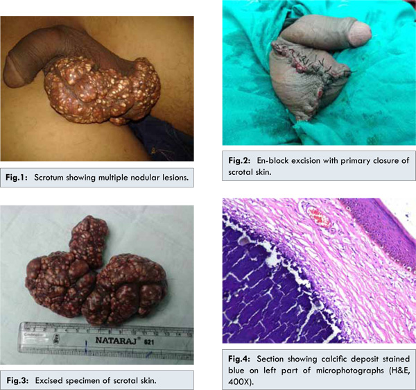
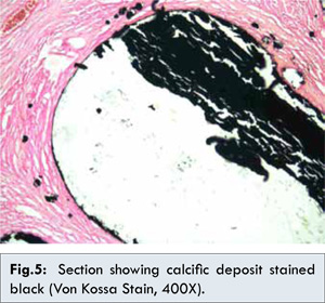
A 45 year old male, referred from a primary health centre, presented with swelling over scrotum for duration of two years. The swelling gradually increased in size. He denied history of scrotal trauma, also had no history of any metabolic, systemic, neoplastic or autoimmune disease. On physical examination, he was healthy and systemic review was normal. On local examination there were multiple nodular lesions involving central part of scrotal skin. There were about 300 nodules grey-brown in colour, hard, non-tender, without any discharge and of various sizes. Penis was not involved [Fig.1]. Clinical diagnosis of multiple sebaceous cysts of scrotum was made. All haematological and biochemical parameters including calcium and phosphorus were within normal limits. En-block excision of involved scrotal lesion of size about 13x8 cm was done with primary closure [Fig.2,3]. Microscopy and pathological analysis of multiple sections showed normal epidermis with multiple small and large deposits of calcium in dermis and subcutaneous tissue. The deposits were not lined by any epithelium [Fig.4]. Von Kossa’s stain for calcium was positive [Fig.5]. The final diagnosis of idiopathic calcinosis cutis of scrotum was made. Patient is under follow-up for the last six months with no recurrence.
Discussion
Idiopathic calcinosis of scrotum was first described by H.M. Lewinsky in 1883 [
2] and then in 1888 by Hutchinson [
3]. However, Shapiro et al. must be given credit for first establishing idiopathic scrotal calcinosis as a distinct entity in 1970 [
4]. It is a rare and benign condition, the exact incidence of which is not known. It commonly occurs in 20 to 40 years of age [
5]. Calcinosis cutis is a term used to describe a group of disorders in which calcium deposits form in the skin. Calcinosis cutis is classified into four major types according to etiology: Dystrophic, Metastatic, Iatrogenic and Idiopathic. Dystrophic and idiopathic varieties are among the few rare types [
6]. Idiopathic calcinosis cutis occurs in the absence of tissue injury or a systemic metabolic disorder. In this condition no etiology is identified and the calcification is localized to one area. Idiopathic calcium deposits have been described in the scrotum, penis, vulva and breast [
7]. Recently a case of extensive Idiopathic scrotal calcinosis was reported by Dombale et al [
8]. Few studies proposed that scrotal calcinosis represents dystrophic calcification of pre-existing structures including epidermal cysts. In the dystrophic form, the serum calcium and phosphorus levels are normal. It may also be observed in connective tissue diseases like scleroderma, dermatomyositis, SLE and secondary to trauma and inflammation [
2,
9,
10].
Treatment of idiopathic calcinosis of scrotum is limited to surgical excision of the affected scrotum with reconstruction. This is generally curative and relapses are rare. The diagnosis of scrotal calcinosis is established by histopathological examination and presence of Von Kossa positive dermal deposits [
9].
In our case, there was no history of connective tissue diseases, trauma and pre-existing scrotal cysts. The biochemical parameters were also normal. Hence, the diagnosis of Idiopathic calcinosis cutis of scrotum fits into our case. The en-block excision of scrotal skin had excellent result and there was no recurrence in a period of 6 months. This case was unique as this is rare variety of calcinosis cutis and there was extensive involvement of scrotum with almost 300 nodules.
Conclusion
Extensive idiopathic calcinosis cutis of scrotum is an uncommon disease with multiple nodules. Excision and primary closure of scrotal skin is the treatment of choice.
References
- Shapiro L, Platt N, Torres-Rodríguez VM. Idiopathic calcinosis of the scrotum. Arch Dermatol. 1970;102(2):199-204.
- Kelten EC, Akbulut M, Colakoglu N, Bayramoglu H, Duzcan SE. Scrotal Calcinosis: is it idiopathic or dystrophic? Aegean Pathol J. 2005;2:4-7.
- Hutchinson J. Illustrations of clinical surgery. Vol 2, Balkiston, Philadelphia, 1882.
- Swinehart JM, Golitz LE. Scrotal calcinosis. Dystrophic calcification of epidermoid cysts. Arch Dermatol. 1982;118(12):985-988.
- Khallouk A, Yazami OE, Mellas S, Tazi MF, El Fassi J, Farih MH. Idiopathic scrotal calcinosis: a non-elucidated pathogenesis and its surgical treatment. Rev Urol. 2011;13(2):95-97.
- Reiter N, El-Shabrawi L, Leinweber B, Berghold A, Aberer E. Calcinosis cutis: Part I. Diagnostic pathway. J Am Acad Dermatol. 2011;65(1):1-12.
- Kanishwar VS, Waghmare RS, Puranik GV. Calcinosis Cutis in the CREST Syndrome. Bombay Hospital J. 2010;52(1):108-110.
- Dombale VD, Basarkod SI, Kotabagi HB, Farheen U. Extensive idiopathic scrotal calcinosis: A case report. J Clin Diagn Res. 2012;6(3):478-479.
- Shah V, Shet T. Scrotal calcinosis results from calcification of cysts derived from hair follicles: a series of 20 cases evaluating the spectrum of changes resulting in scrotal calcinosis. Am J Dermatopathol. 2007;29(2):172-175.
- 10. Dubey S, Sharma R, Maheshwari V. Scrotal calcinosis: idiopathic or dystrophic? Dermatol Online J. 2010;16(2):5.