6go6ckt5b8|3000F7576AC3|Tab_Articles|Fulltext|0xf1ff30f8050000002b03000001000500
6go6ckt5b5idvals|369
6go6ckt5b5|2000F757Tab_Articles|Fulltext
Introduction
Apert syndrome is a rare genetic disorder with an incidence of 15 per million [1]. It has been reported very rarely from India [2]. It is characterised by craniofacial anomalies, craniosynostosis and syndactyly of hands and feet. Other associated anomalies with Apert syndrome include cardiac defects, cleft palate, polycystic kidneys and pyloric stenosis. Early surgical correction of craniofacial anomalies in these cases has been shown to minimise cognitive loss.
These cases pose a challenge to the anaesthesiologists not only because of a difficult airway but also problems like bronchospasm, increased secretion and even a difficulty in securing intravenous access. We report our experience in a case of Apert syndrome posted for endoscopic surgical repair of skull base defect that was a complication of craniosynostosis surgery.
Case Report
A 8-year-old male child, a diagnosed case of Apert syndrome, born of full-term delivery, was referred to our institution for endoscopic correction for CSF rhinorrhoea. This child had mid facial hypoplasia, low lying ears, high arched palate and hypertelorism [Fig.1]. The child achieved normal developmental milestones. No relevant family history was noted.
The patient had history of previous surgeries for syndactyly at the age of 2 years and for craniosynostosis at the age of 3 years [Fig.2]. After craniosynostosis surgery, he had repeated episodes of respiratory tract infection and running nose for which he was treated with antibiotics. He also had a history of meningitis at the age of 7 years. A definitive skull base repair was planned, considering all this. Thorough systemic examination and investigations showed no other associated anomalies.
Nasal packing done by lignocaine 1% with adrenaline 1:100,000 half an hour prior to surgery. Intravenous access was difficult in this child due to limb deformity and 22 G cannula was secured on left limb after multiple attempts. Baseline vitals were recorded. Patient was premedicated by injection glycopyrrolate and injection fentanyl with non-invasive monitors including electrocardiogram, pulse oximetry, and non-invasive blood pressure.
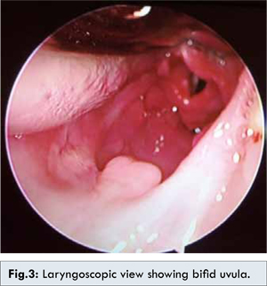
After adequate pre-oxygenation, induction was done by injection propofol 1.5 mg/kg. As anticipated, mask ventilation was difficult because of difficulty in lifting the jaw and inadequate fitting of mask due to mid facial hypoplasia, receding mandible, large tongue and a high arched palate. An oropharyngeal airway was placed to assist ventilation and avoid airway obstruction. After conformation of successful bag and mask ventilation laryngoscopy was done following intravenous injectons of succinylcholine 2 mg/kg and lidocaine 2 mg/kg. Laryngoscopy revealed, high arched palate and bifid uvula [Fig.3]. Intubation was done in first attempt by 5.5 mm cuffed endotracheal tube. After confirmation of endotracheal intubation patient was paralyzed by injection atracurium and placed in 15 degree head up position. Maintenance was done by nitrous oxide 60%, oxygen 40% and propofol infusion to achieve hypotension and creating a bloodless field for endonasal surgery. Intraoperative vitals were stable. After the surgery, Valsalva manoeuvre was applied to check for any leak. After adequate suctioning, the patient was extubated in a deep plane and an oro-pharyngeal airway was placed and oxygenated by face mask. The patient was shifted after return of adequate muscle power and respiration.
Discussion
Apert syndrome is named after French paediatrician, Eugene Apert who first described it in 1906 [3]. These cases are associated with mutation on fibroblast growth factor receptor 2 on chromosome 10q25 [2,4]. It is inherited as an autosomal dominant trait. Apert syndrome accounts for 4.5% of all craniosynosyosis syndromes. Increased incidence is seen with increasing paternal age [5]. Apert syndrome can be diagnosed early in pregnancy, in at risk patients using various methods such as fetoscopy, fetal ultrasound and molecular testing.
Repeated episodes of respiratory tract infection and running nose was suggestive of cerebrospinal fluid rhinorrhea secondary to surgical trauma sustained during craniosynostosis surgery. These patients undergo multiple surgeries including cranial vault remodelling and syndactyly release. CSF rhinorrhea is a complication of cranial vault remodelling which may lead to meningitis as in case of our patient.
Anaesthetic challenges in such cases are mostly related to airway. Bag mask ventilation may be difficult as they have mid face hypoplasia and other facial abnormalities. There is also an increased incidence of bronchospasm in these cases secondary to profuse secretions [6]. There is fusion of C5-C6 cervical vertebra [7].
Incidence of obstructive sleep apnoea is around 50% [8]. We used an airway in our patient after extubation in an attempt to prevent airway collapse after surgery. In our case mask ventilation was difficult but there was no difficulty in intubation which is the case in most patients of Apert syndrome.
Our patient was planned for endonasal correction of skull base defect using fascia lata graft and durafoam. As the nasal cavity is very vascular we kept the patient hypotensive to provide a clear field to the surgeon and minimize blood loss. A Valsalva manoeuvre was given towards end of surgery to check any CSF leak. The patient was extubated in a deep plane as bucking could result in dislodgement of fat graft. An airway placement was done. The patient was monitored till he was fully conscious and then shifted to the ICU. There was no complication and patient was discharged in a healthy condition.
References
- Cohen MM Jr, Kreiborg S. New indirect method for estimating the birth prevalence of the Apert’s syndrome. Int J Oral Maxillofac Surg 1992;21:107-109.
- Sohi BK, Sohi AS. Apert’s syndrome. Indian J Dermatol Venereol Leprol 1980;46:169-172.
- Harper JI. Genetics and genodermatoses. In: Champion RH, Burton JL, Burns D, Breathnach SM, editors. Rook/Wilkinson/Ebling Textbook of dermatology. 6th ed. Oxford: Blackwell Science; 1998. pp. 425-426.
- Wilkie AO, Wall SA. Craniosynostosis: Novel insights into the pathogenesis and treatment. Curr Opin Neurol 1996;9:146-152.
- Fabiola Quintero-Rivera, Caroline D. Robson, Rosemary E. Riess, et al. Apert syndrome: what prenatal radiographic findings should prompt its consideration? Prenat Diagn 2006;26:966-972.
- Elwood T, Sarathy PV, Geiduschek JM, Ulma GA, Karl HW. Respiratory complications during anaesthesia in Apert syndrome. Paediatr Anaesth 2001;11(6):701-703.
- Kreiborg S, Barr M Jr, Cohen MM Jr. Cervical spine in the Apert syndrome. Am J Med Genet 1992;43(4):704-708.
- Bannink N, Nout E, Wolvius EB, Hoeve HL, Joosten KF, Mathijssen IM. Obstructive sleep apnea in children with syndromic craniosynostosis: long-term respiratory outcome of midface advancement. Int J Oral Maxillofac Surg 2010;39(2):115-121.