|
|
|
|
|
A Rare Case of Scrotal Dermoid Cyst Presenting as Third Testis
|
|
|
Bernard Amer, Rajendra Singh Sinam, Vijayendra Singh Kanwar, Naloh Mibang, Kh Somarendra Singh, Md Jawaid Rahman
From the Department of Urology, Regional Institute of Medical Sciences, Imphal-795004, India. |
|
|
|
|
|
Corresponding Author:
|
Dr. Bernard Amer
Email: bernardamer@gmail.com
|
|
|
|
|
|
|
|
|
Received:
19-AUG-2014 |
Accepted:
20-SEP-2014 |
Published Online:
25-OCT-2014 |
|
|
|
|
|
|
|
Abstract
|
|
|
|
A gentleman of 45 years old presented with a painless swelling in the scrotum, of what seemed like a third testis. Clinically, it was a cystic swelling situated at the bottom of the scrotum, separately palpable from both the testis. Computed tomogram (CT) reported it as a cystic swelling arising from the perineum. Fine needle aspiration cytology (FNAC) and excisional biopsy were consistent with dermoid cyst. Its anatomy was accurately defined by CT of pelvis and perineum and guided us in accurate anatomical dissection. Such rare benign lesions should be considered in the differential diagnosis of painless scrotal masses. |
|
|
|
|
|
Keywords :
|
Dermoid Cyst, Genital Diseases, Scrotum, Testis, Pelvis, Humans.
|
|
|
|
|
|
|
|
|
|
|
|
6go6ckt5b8|3000F7576AC3|Tab_Articles|Fulltext|0xf1ff0000060000001303000001000200 6go6ckt5b5idvals|382 6go6ckt5b5|2000F757Tab_Articles|Fulltext Introduction
There are four types of swellings in the perianal region that are more frequently seen namely, epidermoid cyst, dermoid cyst, anal duct/gland cyst and sacrococcygeal teratomas [1]. Dermoid cysts demonstrate squamous epithelium, eccrine glands, and pilosebaceous units. In addition, these particular cysts may develop bone, tooth, or nerve tissue on occasion [2,3]. The incidence of dermoid cyst is unknown, and its rarity may be due to lack of clinical criteria to establish a preoperative diagnosis and the fact that some cases are grouped under mature teratoma [4-8]. Dermoid cyst of the testis should be suspected in a child or adult with a painless testicular mass which has not enlarged for some time. In our case, we encountered an adult patient who presented with a painless testicular swelling for several years which looked like a third testis but later on proved to be a dermoid cyst.
Case Report
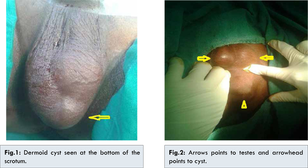
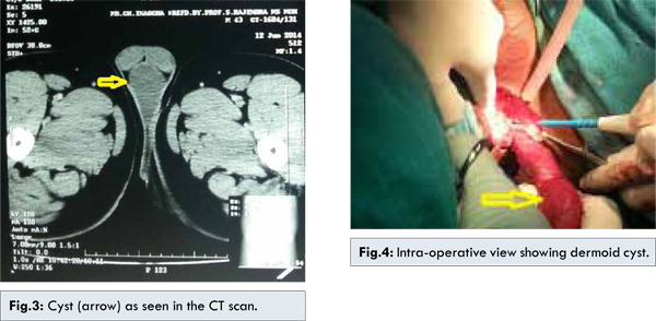
In our out-patient department, a gentleman of age 45 years presented himself to us with complaints of a painless swelling in the scrotum for the past several years. According to him, he could not disclose it or seek medical help earlier due to shame and finally did come to us on the insistence of his wife. We took a thorough history and did a physical examination. On inspection it looked like a third testis situated at the bottom of the scrotum [Fig.1,2]. On palpation it was a soft, cystic swelling clearly separated from the testis as we could palpate the two normal testes in their position. The scrotal skin was normal with no induration or tenderness. There was no impulse on coughing and it did not extend towards the inguinal region. Trans-illumination test was negative indicating that it was neither a clear fluid cyst nor a hydrocele. We asked for a FNAC of the swelling and a CT of pelvis and perineum. Report of FNAC was consistent with dermoid cyst and the CT report was that of a cystic swelling of the scrotum with the apex of the cyst extending towards the perineal body [Fig.3]. There were no other abnormalities such as lymph node enlargements in the retroperitoneum. Given this entire picture, we were confident that this is a benign cystic swelling paving us to forego a tumor marker profiling for testicular tumors. After informing the patient about the possible nature of the swelling and obtaining a written consent, we excised the lesion under spinal anaesthsia and as expected, the histopathological study reported it as a dermoid cyst [Fig.4]. The patient had an uneventful recovery and was discharged from the hospital on the 2nd post-operative day.
A broad spectrum of urological conditions can present with an asymptomatic scrotal swelling; the differential diagnosis of which can sometimes be puzzling [9]. Clinical presentation, imaging and histopathological studies usually differentiate all these types of swelling and guide us in decision making regarding the management. Unusual site of presentation may cause diagnostic dilemma [10]. The cases of midline cyst in the perineum are rare entities and only few cases have been reported in the world literature [11-14]. In fact, one would be considered ‘special’ if he encounters one in his lifetime. This is so because such an encounter is rare due to rareness of the condition itself as well as the patient’s fear of disclosing it. According to the literature, there are mainly four types of perineal swellings. They are epidermal inclusion cyst, dermoid cyst, anal gland/duct cyst and sacrococcygeal teratoma [1]. They grow slowly and are usually asymptomatic until they become inflamed or secondarily infected. They are commonly distributed all over the body but uncommonly reported in the perineum [10]. Dermoid cyst of the testis is filled with sebum and contains sebaceous glands, hair follicles and sometimes sweat and apocrine glands in its fibrous stroma. Bone, cartilage, teeth and brain tissue may be present. The presence of skin appendages differentiates dermoid cysts from epidermal cysts of the testis [15]. Ideal workup of the cyst includes fine needle aspiration for diagnosis and bacterial culture for infected cases. The imaging studies for diagnosis includes sonography, MRI, CT scan and plain radiography. The treatment of perineal dermoid cyst is surgical excision with a careful and meticulous dissection with precaution taken to avoid spillage of the contents and also to avoid injury to vital structures nearby, such as perineal urethra and anal canal [10]. Crankson SJ et al. reported a case of child with testicular dermoid cyst. Their main concern was a solid testicular malignancy [15]. In our case, the patient was an adult and the swelling was cystic, palpable separately from both the testis. Canali R et al. reported a case of scrotal dermoid cyst extenting to the urethra [9]. In our case, the cyst was seen extending towards the perineal body in CT scan which was confirmed during dissection.
Conclusion
A painless, slow growing mass in the scrotum may be anything between benign dermoid cyst to malignant solid tumors. Careful physical examination, use of appropriate imaging techniques and FNAC will often guide us in the diagnosis as well the anatomic details for surgical dissection. A dermoid cyst of the scrotum although very rare should be included in the differential diagnosis of a mass in the scrotum.
References
- Peralta R, Geibel J. Perianal cysts. Available from http://emedicine.medscape.com/article/192071-overview. Accessed on 19th August 2014.
- Mackie RM. Epidermoid cyst. In: Champion RH, Burton JL, Burns DA, et al. (eds): Rook/Wilkinson/Ebling Textbook of Dermatology, Vol. 2, 6th ed. Oxford: Blackwell Science, 1998, pp. 1666.
- Szeremeta W, Parikh TD, Widelitz JS. Congenital nasal malformations. Otolaryngol Clin North Am. 2007;40:97-112.
- Assaf G, Mosbah A, Homsy Y, Michaud J. Dermoid cyst of testis in a five-year-old child. Urology. 1983;2:432-434.
- Burt AD, Cooper G, Mackay C, Boyd JF. Dermoid cyst of the testis. Scott Med J. 1987;32:146-148.
- Maiti S, Chatterjee G, Pal SN, Mukherjee DR. Benign cystic teratoma of the testis. J Indian Med Assoc. 1990;88:287-288.
- Broggi G, Appetito C, di Leone L, Ciprandi G, Menichella P, Broggi M, et al. Dermoid cyst in undescended testis in a nine-year-old boy. Urol Int. 1991;47:110-112.
- Wegner HE, Herbst H, Loy V, Dieckmann KP. Testicular dermoid cyst in an ten-year-old child: case report and discussion of etiopathogenesis, diagnosis and treatment. Urol Int. 1995;54:109-111.
- Canali R, Angelini L, Zhapa MCE, Rigamonti W. Scrotal dermoid extending to the posterior urethra through a corpus cavernosum in a child. Journal of Pediatric Surgery. 2012;47:1618-1621.
- Ali SA, Tahir SM, Memon AS, Dahri AA. Epidermoid inclusion cyst of the perineuma rare case report in a 50 years old male. J Ayub Med Coll Abbottabad. 2009;21(3).
- Takano Y, Yokokawa K, Namiki M, Okuyama A. Perineal Epidermal Cyst. Urol Int. 1994;53(1):53-55.
- Maruyama T, Ueda Y, Suzuk T, Maeda N, Yoshinmoto T, Kondoh N, et al. Epidermoid Cyst of perineo-scrotal region: report of a case. Hinyokika Kiyo. 2004;50(12):8885-8887.
- Machida T, Matsuoka Y, Kobayashi S, Ozeki Z, Ishizaka K, Oka T. A case of giant Perineal Epidermal Cyst: a case report. Hinyokika Kiyo. 2003;49(5):257-259.
- Nagahama K, Sanada S, Mitani T, Yamamuro M, Suzuki T. Giant epidermal cyst in the perineum extending in to the pelvic space in the patient with poly cystic kidney disease: a case report. Hinyokika Kiyo. 2001;47(5):345-348.
- Crankson SJ, Shabra S, Hawashim NA. Dermoid cyst of the testis. Annals of Saudi Medicine. 1997;17(6):634-635.
|
|
|
|
|
|
|
Search Google Scholar for
|
|
|
Article Statistics |
|
Amer B, Sinam RS, Kanwar VS, Mibang N, Singh KS, Rahman MJA Rare Case of Scrotal Dermoid Cyst Presenting as Third Testis.JCR 2014;4:412-415 |
|
Amer B, Sinam RS, Kanwar VS, Mibang N, Singh KS, Rahman MJA Rare Case of Scrotal Dermoid Cyst Presenting as Third Testis.JCR [serial online] 2014[cited 2026 May 15];4:412-415. Available from: http://www.casereports.in/articles/4/2/A-Rare-Case-of-Scrotal-Dermoid-Cyst-Presenting-as-Third-Testis.html |

|
|
|
|
|