|
|
|
|
|
Airway Management in Temporomandibular Joint Ankylosis
|
|
|
Yasemin Burcu Ustun1, Cengiz Kaya1, Yunus Oktay Atalay2, Ersin Koksal1, Ender Cam1
From the 1Department of Anesthesiology and Reanimation and 2Department of Radiology, Outpatient Anesthesia Service; Faculty of Medicine, Ondokuz Mayis University, Samsun, Turkey. |
|
|
|
|
|
Corresponding Author:
|
Dr. Yasemin Burcu Ustun
Email: burcu.ustun@omu.edu.tr
|
|
|
|
|
|
|
|
|
Received:
21-AUG-2014 |
Accepted:
06-OCT-2014 |
Published Online:
05-NOV-2014 |
|
|
|
|
|
|
|
Abstract
|
|
|
|
Temporomandibular joint (TMJ) ankylosis is an important functional pathology that restricts mouth-opening, decreases quality of life, and is associated with facial deformities and orthognathic and orthodontic problems. Airway management in such cases poses a multitude of challenges. This case report pertains to successful airway management in the background of difficult airway. |
|
|
|
|
|
Keywords :
|
Ankylosis, Temporomandibular Joint, Airway Management, Temporomandibular Joint Disorders, Mouth, Humans
|
|
|
|
|
|
|
|
|
|
|
|
6go6ckt5b8|3000F7576AC3|Tab_Articles|Fulltext|0xf1ff1000060000004303000001000100 6go6ckt5b5idvals|386 6go6ckt5b5|2000F757Tab_Articles|Fulltext Introduction
Temporomandibular joint (TMJ) ankylosis occurs as a result of the fusion between the skull base and the mandibular condyle. Although trauma is the most important etiological factor, otitis media, mastoiditis, or hematogenous infections can induce TMJ ankylosis [1,2]. Rheumatoid arthritis, Paget’s disease, ankylosing spondylitis, pseudohypoparathyroidism, psoriasis, and burns can also play a role in its etiology [1,3]. Ankylosis of the TMJ cannot be detected during the early stages of childhood. In ankylosis of the TMJ, problems such as speech disorders, difficulty with mastication, bad oral hygiene, and widespread caries affect the physical and psychological development of the patient [2,3]. Herein, we aim to present airway management of a patient who will be undergoing a series of operations performed by a plastic surgeon.
Case Report
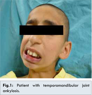
A 13-year-old boy weighing 35 kg was presented to the surgery unit for TMJ arthroplasty. He had previously been operated on 8 years earlier for TMJ ankylosis that had developed following an ear infection. The patient could take only fluids, and his weight percentile was lower than his peers. Mouth-opening (<1 cm) was restricted on the preoperative airway evaluation [Fig.1].
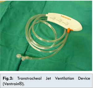
Routine monitoring (pulse oximeter, indirect blood pressure, electrocardiogram and capnography) was performed in the operating room. To alleviate anxiety, intravenous (IV) midazolam (0.03 mg/kg) was administered and remifentanil IV infusion (0.01 mcg/kg/min) was started. A complete set of resuscitation equipment was prepared, i.e., nasal airway (6-7 mm) and transtracheal jet ventilation device (Ventrain®, Eindhoven, The Netherlands, Fig.2). Our hospital’s pediatric-sized fiberoptic device was broken and could not be used. For anesthetic induction, a total dose of 3 mg/kg propofol was titrated. Since mask ventilation was easily performed, intubation was not attempted. Following achievement of adequately deep anesthesia, the ENT team proceeded with the tracheostomy operation, and a tracheostomy cannula was placed 15 minutes later. After airway patency was ensured, rocuronium (0.6 mg/kg IV) was injected and the patient was connected to a ventilator. Anesthesia was maintained using oxygen and sevoflurane. A plastic and reconstructive surgeon performed gap arthroplasty on the right temporomandibular joint. At the end of the surgery, the neuromuscular block was reversed using sugammadex (3 mg/kg IV) and the patient was transferred to the post-anesthesia care unit (PACU).
In TMJ, ankylosis intubation with conventional methods is almost impossible. Blind nasal intubation, fiber optic-guided intubation, retrograde intubation, and tracheostomy are alternative interventions [4]. Although awake nasal intubation is a safe alternative, its application in children is challenging. Blind nasal or fiberoptic-guided nasal intubation is included in the algorithm, but laryngospasm [5] and soft tissue bleeding [6] can further complicate intubation. In our patient, since recurrent surgical operations were planned, in consultation with the surgical team, open surgical tracheostomy was preferred. Since our pediatric fiberoptic device was broken and we had no blind nasal intubation experience with children, we could not use these alternatives of the algorithm. It was decided pre-operatively that in case of failure with mask ventilation, switching to transtracheal jet ventilation will be done. Transtracheal jet ventilation is applied using high-ventilation velocities, which provides tidal volumes equal to or lower than the patient’s end tidal alveolar dead space. To emergently provide a patent airway, with the aid of an intracath (20G), the trachea is entered and transtracheal jet ventilation is applied, which can be life-saving in patients whose airway patency cannot be ensured otherwise. With Ventrain®, airway patency can be safeguarded without any need for laryngoscopy, and respiratory support can then be provided with an intratracheal catheter [7-9]. Therefore, we thought to include Ventrain® in our treatment protocol. Mask ventilation was easily performed in our patient, so percutaneous transtracheal jet ventilation was not required. Airway patency was ensured by surgically placing a tracheostomy cannula. In our case, according to the algorithm concerning management of a difficult airway, before the invasive intervention a local laryngeal nerve block and application of topical anesthesia on the upper airway could allow blind nasal intubation.
In conclusion, for patients in whom complications of intubation are anticipated, detailed preoperative preparation can decrease morbidity and mortality rates. We think that a Ventrain® device should be available during preoperative preparation.
References
- Regev E, Koplewitz BZ, Nitzan DW, Bar-Ziv J. Ankylosis of the temporomandibular joint as a sequela of septic arthritis and neonatal sepsis. Pediatr Infect Dis J. 2003;22:99-101.
- Kumar R, Hota A, Sikka K, Thakar A. Temporomandibular joint ankylosis consequent to ear suppuration. Indian J Otolaryngol Head Neck Surg. 2013;65:627-630.
- Chidzonga MM. Temporomandibular joint ankylosis: review of thirty-two cases. Br J Oral Maxillofac Surg, 1999;37:123-126.
- Enterlein G, Byhahn C. Practice guidelines for management of the difficult airway: update by the American Society of Anesthesiologists Task Force. Anaesthesist. 2013;62:832-835.
- Kawasaki T, Sata T, Kawasaki C, et al. Airway management of a child with temporomandibular joint ankylosis following otitis media. Anaesthesia. 2002;57:294-295.
- TK Sahoo, Y Patil, Patel RD, Dewoolkar LV. Anaesthetic management of a child with temporomandibular joint ankylosis with extrahepatic portal vein obstruction for ankylosis release. The Internet Journal of Anesthesiology. 2008;16:1.
- Ahmad Y, Turner MW. Transtracheal jet ventilation in patients with severe airway compromise and stridor. Br J Anaesth, 2011;106:602.
- Hamaekers AE, Borg PA, Enk D. Ventrain: an ejector ventilator for emergency use. Br J Anaesth. 2012;108:1017-1021.
- Ross-Anderson DJ, Ferguson C, Patel A. Transtracheal jet ventilation in 50 patients with severe airway compromise and stridor. Br J Anaesth. 2011;106:140-144.
|
|
|
|
|
|
|
Search Google Scholar for
|
|
|
Article Statistics |
|
Ustun YB, Kaya C, Atalay YO, Koksal E, Cam EAirway Management in Temporomandibular Joint Ankylosis.JCR 2014;4:428-430 |
|
Ustun YB, Kaya C, Atalay YO, Koksal E, Cam EAirway Management in Temporomandibular Joint Ankylosis.JCR [serial online] 2014[cited 2026 May 27];4:428-430. Available from: http://www.casereports.in/articles/4/2/Airway-Management-in-Temporomandibular-Joint-Ankylosis.html |

|
|
|
|
|