|
|
|
|
|
Osteogenic Sarcoma of Mastoid Bone
|
|
|
mixing viagra and weed mixing adderall and weed open
From the Department of Head & Neck Surgical Oncology, Regional Cancer Centre (RCC), Trivandrum, Kerala, India. |
|
|
|
|
|
Corresponding Author:
|
Dr. Santhosh Kumar N
Email: dr.santhosh82@gmail.com
|
|
|
|
|
|
|
|
|
Received:
08-JUL-2014 |
Accepted:
18-AUG-2014 |
Published Online:
05-SEP-2014 |
|
|
|
|
|
|
|
Abstract
|
|
|
|
Osteosarcoma is the most common primary malignant tumour of the bone. The involvement of the craniofacial bones in osteosarcoma is relatively rare. The mandible and the maxilla are the most commonly affected bones of the head. We report a rare low-grade osteogenic sarcoma of the mastoid part of the temporal bone. |
|
|
|
|
|
Keywords :
|
Osteosarcoma, Mastoid, Maxilla, Temporal bone, Bone Neoplasms, Head and Neck Neoplasms.
|
|
|
|
|
|
|
|
|
|
|
|
6go6ckt5b8|3000F7576AC3|Tab_Articles|Fulltext|0xf1ff6cf0050000001c03000001000800 6go6ckt5b5idvals|361 6go6ckt5b5|2000F757Tab_Articles|Fulltext Introduction
Sarcomas are malignant neoplasms originating from mesodermal tissues and constitute less than 1% of body’s tumors. Osteosarcomas are the most common primary malignant tumor of the bone. The long bones of the limbs, especially the distal end of the femur and the proximal end of the tibia are most commonly involved. Osteosarcoma of the craniofacial bones is relatively rare and represents less than 10% of all osteosarcomas. The most commonly affected bones of the head are the mandible, followed by the maxilla. Osteosarcomas of extra-gnathic craniofacial bones are very rare, constituting less than 2% of all osteosarcomas. Osteosarcomas account for approximately 1% or less of all head and neck cancers [1-3].
Case Report
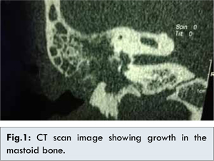
We report a case of 37 year old female who presented to medical college, Trivandrum with history of decreased hearing and tinnitus of the right ear for 6 months. On examination, there was a red, firm mass filling the right external auditory canal. The pure tone audiometry and tuning fork tests were suggestive of moderate conductive hearing loss. High resolution CT scan was showing soft tissue density in the right external auditory canal involving bony and cartilaginous part extending to the middle ear with ossicular erosion [Fig.1]. There was also erosion of the scutum and soft tissue density in the mastoid air cells. The patient was diagnosed to have cholesteatoma and underwent modified radical mastoidectomy with informed consent. Intraoperatively it was found to have red, hard mass filling the mastoid antrum, mastoid air cells, external auditory canal and destroying the posterior canal wall. The tissue was sent for histopathological examination and was showing low grade osteosarcoma. Then she was referred to our institute for further management.
The histopathological slides were reviewed and confirmed diagnosis of low grade osteosarcoma [Fig.2,3]. The haematological and biochemical parameters were normal. On evaluation, technetium 99 MDP scan was showing a hot spot over the right external auditory canal corresponding to the primary site. Radiological evaluation of the brain and thorax were normal, but MRI was showing a small T2 hypointense enhancing nodular lesion.
The case was discussed in tumour board, because of the small residual lesion and the patient’s refusal for another surgery; she was planned for treatment with external beam radiotherapy.
Discussion
Osteogenic sarcoma of the craniofacial bone is very rare. It occurs predominantly in the mandible followed by the maxilla. Mastoid bone as site of osteogenic sarcoma is a rare presentation. Osteosarcomas of extragnathic craniofacial bones constitute less than 2% of all osteosarcomas, and less than 0.5% of all primary bone tumors.
Nora et al. reported a series of 21 patients with osteosarcomas of extragnathic craniofacial bones. They revised the archives of Mayo Clinic containing nearly 7000 bone tumors, including 1000 osteosarcoma. Among them only 21 cases were affecting extragnathic craniofacial bones. The usually involved site among the cranial bones was the occipital bone. Only one of these cases was located at the temporo-sphenoid region [2]. Salvatti et al. also presented 19 cases of osteosarcoma of the skull, only one of which was located at the temporo-parietal bone region [3]. Seely and Gates reported two cases of parosteal osteosarcoma arising from mastoid bone [4]. Kumar et al. also reported eight patients with parosteal osteosarcoma of cranial bones, only two of them were arising from the mastoid region [5]. Brusic SK et al. reported a case of 75-year-old female with osteosarcoma of the mastoid process with history of radiation therapy after total parotidectomy and radical neck dissection in treatment of mucoepidermoid carcinoma of the parotid gland 12 years before the presentation [6]. Hsieh et al. reported a case of parosteal osteosarcoma of the mastoid bone following radiotherapy for nasopharyngeal carcinoma [7].
Secondary osteosarcomas occur more commonly in skull bones. The most common predisposing factors being Paget’s disease or exposure to radiation. Osteosarcoma is a rare complication of radiation therapy and usually occurs after a long latent period. Hereditary retinoblastoma, history of fibrous dysplasia, or trauma constitute other predisposing features [8]. The present case appears to have developed de novo; as no history of any predisposing factors to be elicited from this patient.
The most common presenting symptom of osteosarcoma in the head and neck region is swelling [9]. Pain was the presenting complaint in approximately 50% of the patients [10]. Pathological fractures can occur in cases of large sarcomas. Sensory neural abnormalities may occur in cases where the lesion involves the course of peripheral nerves. Involvement of temporo-mandibular joint or muscles of mastication may cause trismus [12,13]. In contrary to the above findings, our patient presented with hearing loss and tinnitus which are more in favour of chronic ear disease. Therefore, the primary physicians thought in favour of cholesteatoma.
Parosteal osteosarcomas with typical radiological appearance of sessile, densely ossified surface growth with radiating bone spicules that blend with surrounding soft tissue are usually low grade lesions and en bloc resection is curative in most cases. Radiographically, osteosarcoma usually produces poorly defined, irregular destructive bone lesion interspersed with radioopaque material [14]. Osteosarcoma of the head and neck are considered by most clinicians to be distinct from osteosarcoma that arise in the long bones. It is reported to affect older patients and follows a different clinical pattern [8].
Masoid osteosarcomas remain enigmatic in many ways due to the difficulties related to their diagnosis and treatment especially en-bloc resection. The optimal treatment is surgery. Complete excision with a true en-bloc resection with clear margins is a cornerstone in the treatment of osteosarcoma in the head and neck region. However, due to the presence of many critical nervous and vascular structures in this region, complete excision in an en-bloc fashion is often extremely difficult. Adjuvant radiotherapy should be considered for those with close or positive margins. The role of adjuvant chemotherapy is ill-defined. The likelihood of cure is approximately 60-70%. Mendenhall WM, reported that the prognosis of patients with osteosarcoma were statistically better with the absence of paraesthesia, smaller tumour size, younger age, adequacy of surgical removal and a more differential histological grade of the lesion [15]. Fergusen et al. reported that surgery is the vital modality for treating osteosarcoma and the adjuvant chemotherapy play an important role in the control of subclinical metastatic disease. They also suggested that for the patients with complete surgical excision is not possible, the addition of radiation therapy allows better local control [16].
Conclusion
Osteosarcoma of mastoid bone is an uncommon tumor with unique therapeutic challenges by virtue of its anatomical location, lack of standardized treatment protocols, and poor tolerability to conventional treatment options, and hence needs to be addressed differently from extremity osteosarcomas. Because of the relative paucity of patients with head and neck osteosarcoma, multi-institutional collaborative studies are required to devise future therapeutic strategies for this tumor.
References
- Malawer MM, Link MP, Donaldson SS. Sarcomas of bone. In: DeVita VT Jr, Hellman S, Rosenberg SA, eds. Cancer. 5th edn. Philadelphia, PA: Lipincott-Raven,1997:1789-1852.
- Nora FE, Unni KK, Pritchard DJ, et al. Osteosarcoma of extragnathic craniofacial bones. Mayo Clin Proc 1983;58:268-272.
- Salvati M, Ciappetta P, Raco A. Osteosarcomas of the skull. Clinical remarks on 19 cases. Cancer 1993;71:2210-2216.
- Seely DR, Gates GA. Parosteal osteogenic sarcoma of the mastoid bone. Ann Otol Rhinol Laryngol 1997;106:729-732.
- Kumar R, Moser RP Jr, Madewell JE, Edeiken J. Parosteal osteogenic sarcoma arising in cranial bones: clinical and radiologic features in eight patients. Am J Roentgenol 1990;155:113-117.
- Brusic SK, Pusic M, Cvjetkovic N, Karnjus R, Candrlic B, Kukuljan M, et al. Osteosarcoma of the mastoid process following radiation therapy of mucoepidermoid carcinoma of the parotid gland--a case report. Coll Antropol. 2012;36 Suppl 2:223-225.
- Hsieh ST, Guo YC, Tsai TL, Li WY, Lin CZ. Parosteal osteosarcoma of the mastoid bone following radiotherapy for nasopharyngeal carcinoma. J Chin Med Assoc 2004; 67(6):314-316.
- Ogunlewe MO, Ajayi OF, Adeyemo WL, Ladeinde AL, James O. Osteogenic sarcoma of the jaw bones: A single institution experience over a 21-year period. Oral Surg Oral Med Oral Pathol Oral Radiol Endod 2006;101:76-81.
- Nissanka EH, Amaratunge EA, Tilakaratne WM. Clinicopathological analysis of osteosarcoma of jaw bones. Oral Dis 2007;13:82-87.
- Mark R J, Sercarz JA, Tran L, Dodd LG, Selch M, Calcaterra TC. Osteogenic sarcoma of the head and neck. The UCLA experience. Arch Otolaryngol Head Neck Surg 1991;117:761-766.
- Tanzawa H, Uchiyama S, Sato K. Statistical observation of osteosarcoma of the maxillofacial region in Japan. Analysis of 114 Japanese cases reported between 1930 and 1989. Oral Surg Oral Med Oral Pathol 1991;72:444-448.
- Adekeye EO, Chau KK, Edwards MB, Williams HK. Osteosarcoma of the jaws – A series from Kaduna, Nigeria. Int J Oral Maxillofac Surg 1987;16:205-213.
- Bianchi SD, Boccardi A. Radiological aspects of osteosarcoma of the jaws. Dentomaxillofac Radiol 1999;28:42-47.
- Daw NC, Mahmoud HH, Meyer WH, Jenkins JJ, Kaste SC, Poquette C A, et al. Bone sarcomas of the head and neck in children: The St Jude Children’s Research Hospital experience. Cancer 2000;88:2172-2180.
- Mendenhall WM, Fernandes R, Werning JW, Vaysberg M, Malyapa RS, Mendenhall NP. Head and neck osteosarcoma. Am J Otolaryngol 2011;32:597-600.
- Fergusen WS, Goorin AM. Current treatment of osteosarcoma, Cancer Invest 2001;19(3):292-315.
|
|
|
|
|
|
|
Search Google Scholar for
|
|
|
Article Statistics |
|
Kumar N S, Mathew Iype E, Thomas S, K Jayasree , Ganesan SOsteogenic Sarcoma of Mastoid Bone.JCR 2014;4:334-337 |
|
Kumar N S, Mathew Iype E, Thomas S, K Jayasree , Ganesan SOsteogenic Sarcoma of Mastoid Bone.JCR [serial online] 2014[cited 2026 May 14];4:334-337. Available from: http://www.casereports.in/articles/4/2/Osteogenic-Sarcoma-of-Mastoid-Bone.html |

|
|
|
|
|