Introduction
Fine-needle aspiration biopsy (FNAB), which is applied often in head-neck clinics, is a cheap and effective diagnosis process. While it is often applied directly, it can also be done with the help of imaging methods [
1,
2]. In addition to the usual minor complications such as pain, irritation, rash around the area where biopsy is made, major complications that threaten life such as carotid hematoma might also be seen rarely [
3,
4]. We hereby report a carotid hematoma case which emerged after a fine-needle aspiration biopsy.
Case Report
25-year old female patient consulted our clinic with the complaints of throat-ache and neck swelling. Upon ear-nose-throat examination of the patient, a semi-firm consistent, mobile mass with a 1x1cm size was detected at the right level-IV on the neck. The patient stated that this swelling on the neck emerged 2 months ago. The patient’s blood count, C reactive protein (CRP), sedimentation and other biochemical blood tests were within normal limits. The patient’s ultrasonography report noted “a 10 mm sized, round shaped, heterogeneous nodular-cystic view, with a necrotic view inside, which had tick walls and no bleeding on its periphery with Doppler,” on the right-side level IV of the neck. For the purposes of cytopathological assessment, the fine-needle aspiration biopsy process was planned.
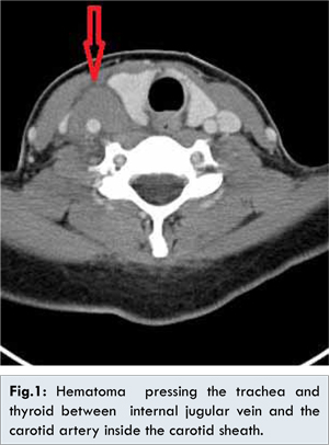
The area above the mass was cleaned with a 10% povidone iodic solution, and 22 -gauge needle was inserted into the 1x1 cm sized mass, which was palpated at Level-IV on the right side of the neck. During the aspiration, firstly around 2 cc. green serous fluid came out, and immediately afterwards high-pressure hemorrhagic fluid was aspirated. We applied digital pressure in area where the needle made the penetration. Within minutes, swelling appeared which filled the right-side levels III and IV of the patient’s neck. This was accompanied by pain in neck and crepitation. Considering carotid hematoma, an urgent neck ultrasound was done, and a view matching hematoma was detected on the right main carotid artery and jugular vein. In order to assess the mediastinum extension of the hematoma size precisely, computed tomography scan was planned. The neck CT result reported that “hematoma around the right carotid artery, that reached 12 mm size at its thickest point, and had an average of 70 HU density; right-lobe of thyroid and trachea were pushed to the left; in the thyroid inferior vicinity, and in connection with the anterior jugular vein, there was a view matching the active contrast extravasation” [Fig.1].
The patient was hospitalized with the diagnosis of carotid hematoma. The pressure bandage was applied on carotid hematoma. The patient was immobilized with elevated head and was closely monitored. Afterwards, the patient’s full neurological examination was made, and no deficit was detected. Endoscopic larynx examination revealed normal vocal cord movements. Following the 24 hour scrutiny, full blood test and neck ultrasound were made on the patient. In the full blood test results, no fall in hemoglobin, hematocrit and reduction of the blood cells were detected. The neck ultrasound report noted that there was a shrinking (at its widest point 12 x 20 mm) hematoma inside the carotid sheath.
In the 3rd day of the scrutiny of the patient in our service, there were significant decrease in the complaints of the patient, and the follow-up ultrasound report noted “a minimal hematoma inside the carotid sheath.” The patient was discharged from the hospital with advice to visit polyclinic for follow-up examinations. The patient’s cytopathology results reported findings which were “matching chronicle inflammation”. During the 2nd week follow-up examination of the patient, the swelling on the right side of the neck was completely resorbed. In addition no mass lesion was detected on the area, and the patient’s case was followed closely.
Discussion
Fine-needle aspiration biopsy (FNAB) is an often used method, which enables us to make cytological examination by applying 20, 22, 25-gauge needles and thinner needles [
1,
2]. FNAB is used quite often because it is safe, cheap and has a high diagnostic value. For the neck masses in general and the thyroid in particular, FNAB’s sensitivity is estimated around 83% and its specificity around 92% [
5]. It can be performed quite easily and without much finanacial burden on patient [
1,
3,
6]. If enough amounts can be aspirated, the results can be obtained within 24 hours after the cytologist performs the procedure.
FNAB often holds an important place in the head-neck surgical clinics in approaching the neck masses, particularly the thyroid and lymph nodules. In many clinics it became a routine process for patients who are diagnosed with having neck masses [
6,
7]. In Western countries, 86 percent of the otorhinolaryngologists carry out an average of 4.7 FNAB operations a month [
1]. At head and neck clinics, one applies FNAB to many palpable masses without sonograhic view. Nevertheless, in cases of masses which are non-palpable and whose vascular structure nearby are known, the FNAB is applied together with sonographic viewing [
2,
7].
The complications which might occur during FNAB’s application to the head-neck region are rare and mostly insignificant. The most common ones are pain and irritation, and minor hematoma [
3,
8]. Life-threatening complications such as the massive hematoma which pressures the airway, post-hematoma neuritis, pseudoaneurysm, carotid artery hematoma, secondary hemangioma, acute temporary thyroid, gland edema, infection, recurring nerve paralysis, vasovagal reaction, tracheal perforation, dysphagia, emergence of sinus along the needle tract, change in the volume of nodule, post-aspiration growth of thyrotoxicosis are also emphasized in the literature [
3,
4]. After the application of the FNAB, it is possible to observe the growth of the hematoma within hours or days. However, many of these are asymptomatic cases, where the hematoma spontaneously reabsorbs within days.
The growth of the carotid artery hematoma after the application of FNAB is rarely stated in the literature. Yet, it can rapidly damage safety of the airway, it is a dangerous major complication [
3,
4,
8]. Carotid artery hematoma occurs as a result of the ponding of blood into the carotid sheath or the neck tissues, which is generated by the increase in pressure caused by damaging of the carotid artery sheath and its wall during the FNAB process. This situation can damage the respiration, neural impulses, arterial and venous feeding, and can reach to a life-threatening position within a short time. While the carotid artery hematoma may grow after the application of FNAB, it can also happen during the internal jugular vein catheterization [
9,
10].
If the patient who underwent the FNAB suffers from pain, sensitivity and mass effect within minutes after the process, then the possibility carotid artery damage should be primarily considered [
3,
4]. If, in addition to the symptoms, the anatomical similarity is also observed, then first of all fingers pressure must be applied. In such a case, within a short time, the hematoma should be assessed by using the sonographic viewing. Through hematoma pressure, a tracheal push might emerge. In the case of our patient, carotid hematoma had pushed the trachea along with the thyroid tissue towards the left; however, no respiratory distress was observed on our patient during this time. The hematomas that limit themselves and which do not cause respiratory distress, in most cases are resorbed spontaneously [
3,
4]. The patient must be kept under close scrutiny, and efforts should made to keep the coagulation profile within the normal boundaries. Through sonograhic monitoring, the hematoma reabsorbtion should be observed [
3,
4]. However, if the patient suffers from respiratory distress, the hematoma drainage should be carried out while maintaining hemodynamics.
The most common reason for the occurrence of the carotid artery hematoma after the application of the FNAB process is the anatomical vascular proximity. For this reason if the contours of the mass cannot be determined precisely through palpation, then by taking into consideration the vascular anatomical proximity, the FNAB process should be recommended together with the sonographic viewing. However, despite these, there are examples in the literature, which point to the occurrence of the carotid hematoma after the application of the FNAB [
3,
4]. Presence of factors like bleeding disorders and hypertension highly increase the risk of the carotid hematoma after FNAB. In the cases of such patients, the International Normalization Rate (INR) should be carefully checked and followed [
11].
Conclusion
Although the application of the FNAB on the head-neck region is quite a safe method, we believe that being cognizant of the major complications, assessing the benefit-damage ratio for the patient properly will further reduce the already-low morbidity rate in these processes. By presenting the case of growth of the carotid hematoma in a patient, we wanted to draw attention to the major complications of this method as well.
References
- Cannon CR, Replogle WH. Fine-needle aspiration: survey of clinical utility. Otolaryngol Head Neck Surg. 2000;123:563-565.
- Braga M, Cavalcanti TC, Collaco LM, Graf H. Efficacy of ultrasound-guidedfine- needle aspiration biyopsy in the diagnosis of complex thyroid nodules. J Clin Endocrinol Metab. 2001;86:4089-4091.
- Polyzos SA, Anastasilakis AD. Clinical complications following thyroid fine-needle biopsy: a systematic review. Clin Endocrinol. 2009;71:157-165.
- Anastasilakis AD, Polyzos SA, Nikolopoulos P. Subendothelial carotid hematoma after fine-needle aspiration biopsy of a solitary thyroid nodule. J Ultrasound Med. 2008;27(10):1517-1520.
- Gharib H, Goellner JR. Fine-needle aspiration biopsy of the thyroid: an appraisal. Ann Intern Med 1993;118:282-289.
- Noordzij JP, Goto MM. Airway compromise caused by hematoma after thyroid fine-needle aspiration. Am J Otolaryngol. 2005;26(6):398-399.
- Ronald G. Amedee, Nina R. Dhurandhar. Fine-Needle Aspiration Biopsy. Laryngoscope. 2001; 111:1551-1557.
- Hor T, Lahiri SW. Bilateral thyroid hematomas after fine-needle aspiration causing acute airway obstruction. Thyroid. 2008;18(5):567-569.
- Teichgräber UK, Benter T, Gebel M, Manns MP. A sonographically guided technique for central venous access. Am J Roentgenol. 1997;169:731-733.
- Karakitsos D, Labropoulos N, De Groot E, Patrianakos AP, Kouraklis G, Poularas J, et al. Real-time ultrasound-guided catheterisation of the internal jugular vein: a prospective comparison with the landmark technique in critical care patients. Crit Care. 2006;10(6):R162.
- Tsilchorozidou T, Vagropoulos I, Karagianidou C, Grigoriadis N. Huge intrathyroidal hematoma causing airway obstruction: a multi disciplinary challenge. Thyroid. 2006;16:795-799.