Introduction
Serous ovarian cystadenocarcinomas account for 25% of serous tumors. They account for the largest proportion of malignant ovarian tumors [
2], representing over 50-80% of all malignant epithelial ovarian tumors [
3]. The prevalence peaks around the 6th to 7th decades of life [
4]. Ovarian neoplasm usually presents with a pelvic mass with or without presence of ascites depending upon the stage of the disease. Malignancy is strongly suspected if the mass has complex solid cystic appearance with raised levels of tumor markers [
1]. We hereby present a case of giant papillary cystadenocarcinoma of ovary in 58 year old female with omental metastasis.
Case Report
58 year old female presented in outpatient department with chief complaints of abdominal distension, pain abdomen especially in left lumbar region since last 1 year. Pain was dull aching in nature and continuous which used to relieve with oral analgesics. Patient also complained of occasional breathlessness and fullness in abdomen. Breathlessness was precipitated in supine position which was relieved in sitting position. She also complained of loss of appetite and significant weight loss. There was no history of bleeding per vaginum. There was no previous history of any major medical or surgical illness.
On examination, general condition was fair with stable vital parameters. Per abdomen examination showed mass of 22 weeks size with irregular surface, with restricted mobility. Ascites was present and positive fluid thrill was noted. On per speculum examination, flushed cervix and vagina was seen. Per vaginum examination showed bosselated mass with irregular surface of 22 week size with restricted mobility arising from the pelvis but uterus was not felt separately.
Patient had gone to rural hospital for the same complaints where ultrasound was done which was suggestive of ill-defined heterogenous soft tissue of size 13x7x9 cms seen arising from the right pole of pelvic region and extending into abdomen. Lesion showed area of necrosis but no area of focal calcification. Similar lesion was seen on the left pelvic region with 11x7x10 cms. Left ovary was not seen separately. Presence of ascites was appreciated. CT abdomen and pelvis revealed lobulated abdo-pelvic mass most likely arising from ovary suggestive of ovarian neoplasm with enlarged lymph nodes with possible omental metastasis and ascites. Ascitic fluid tapping revealed no evidence of malignancy. FNAC report was suggestive of epithelial malignancy highly suggestive of serous papillary cystadenocarcinoma.
Patient was taken for exploratory laparotomy. A midline vertical incision was taken over lower abdominal wall. Abdomen was opened in layers which had substantial fibrosis in the tissues. Bilateral complex vascular ovarian tumor of size 8x10 cms each was seen on both the sides with adhesions present between the bowel, uterus, omentum and the tumor. Pedicle of ovarian tumour was clamped and cut and ligated by Vicyl No. 1-0. Adhesiolysis was done between uterus and omentum. Bilateral infundibular pelvic ligaments were clamped, cut and ligated by Vicryl 1-0. Bilateral uterine artery and bilateral utero-sacral ligaments clamped, cut and ligated by Vicryl 1-0 individually. Vault was sutured with Vicryl 1-0 by continous interlocking sutures. Bilateral lymph nodes of common iliac, internal iliac, external iliac and obturator nodes were removed and omentectomy done. Haemostasis achieved and abdomen was closed in layers with suction drain in situ [Fig.1].
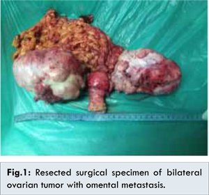
Histo-pathological report revealed papillary serous cystadenocarcinoma of bilateral ovaries. Involvement of 1 out of 10 lymph nodes on right side and 1 out of 9 lymph nodes on left side was present. Metastasis to omentum was present [Fig.2]. Patient underwent chemotherapy for the same. 2 cycles of paclitaxel 250 mg and carboplastin 400 mg were given. On follow up, wound had healed well and patient is stable.
Discussion
Serous papillary cystadenocarcinoma is one of the challenging situation in surgical oncology. It is one of the most common tumors of ovary which generally presents at later stages due to its aggressive nature and delay in the diagnosis and presentation. Macroscopically serous cystadenocarcinoma appear as multilocular cystic ovarian tumor with papillary projections. Psammomatous bodies, characteristic of papillary cystadenocarcinoma of ovary may be present in 30% of cases on histology. Cancer of the ovary is bilateral in 25% [
5].
On ultrasound, papillary serous cystadenocarcinoma is more heterogenous in appearance than serous cystadenoma. Papillary projections, thick septations and solid component amongst the ovarian mass can also be appreciated on ultrasound. Colour Doppler may be helpful in confirming the vascularity of the solid part of the tumor but advantage of using resistivity index and pulsatility index is still doubtful. Ascites seen on ultrasound, points towards presence of peritoneal metastasis. CT scan for ovarian tumors is helpful in staging of disease as it gives information pre-operatively about lymphadenopathy, peritoneal deposits and distant metastasis.
MRI is the modality of choice in the characterization of ovarian malignancy and in the detection of lymphatic, peritoneal, and distant metastases, both for preoperative planning and post treatment follow up. The cystic components are high T2, low T1 signal, unless there has been intra-lesional hemorrhage (where as mucinous cystadenocarcinoma, where there is typically slightly increased T1 signal of the cystic component). Solid malignant components demonstrate intermediate T1 and T2 signal, restricted diffusion, and gadolinium enhancement. DWI (diffusion weighted imaging) is useful for detection of distant metastases [
6,
7].
The abdominal lumps at the perimenopausal and postmenopausal women should be investigated thoroughly so as to know the nature or behavior of the tumor. Facility of frozen section is mandatory if the diagnosis is not certain. Except for tumors with stage Ia, chemotherapy with cisplatin and paclitaxel is a must in adjuvant set up. As our patient was stage IIIc, we had done bilateral oopherctomy, total hysterectomy with lymph node dissection and omental resection followed by chemotherapy. This mode of management helped our patient to overcome her cancer and she went home in a relatively healthy state.
Conclusion
Management of serous papillary cystadenocarcinoma is challenging. Complete surgical resection without spillage of tumor with adjuvant therapy is the treatment of choice. Ultrasound guided fine needle aspiration cytology and peritoneal fluid sampling help in pre-operative diagnosis of the pathology. MRI is the ideal investigation for pre-operative staging of the disease but exploratory laparotomy with post-operative histo-pathology report is the most reliable for final staging of the disease.
References
- Katke RD, Gadekar S, Shaikh A, Aghav B. Bilateral serous cystadenofibroma of ovary with paratubal cyst: a rare case report. Int J Reprod Contracept Obstet Gynecol. 2014;3:764-766.
- Hamm B, Forstner R, Beinder E, et al. MRI and CT of the Female Pelvis. Springer Verlag, Berlin Heidelberg;2007.
- Kawamoto S, Urban BA, Fishman EK. CT of epithelial ovarian tumors. Radiographics. 1999;19:S85-102.
- Ros PR, Mortele KJ. CT and MRI of the abdomen and pelvis, a teaching file. Lippincott Williams & Wilkins, Philadelphia, 2006.
- Cotran RS, Kumar V, Collins T, Robbins Pathologic Basis of Disease, W.B. Sauders, Philadelphia, Pa, USA, 1999.
- Outwater EK, Huang AB, Dunton CJ, Talerman A, Capuzzi DM. Papillary projections in ovarian neoplasms: appearance on MRI. J Magn Reson Imaging. 1997;7(4):689-695.
- Imaoka I, Wada A, Kaji Y, Hayashi T, Hayashi M, Matsuo M, et al. Developing an MR imaging strategy for diagnosis of ovarian masses. Radiographics. 2006;26(5):1431-1448.