Introduction
Gallstone ileus (GI) is characterized by occlusion of the intestinal lumen as a result of one or more gallstones. According to reports from the 1990s, GI is a rare complication of gallstones that occurs in 1%-4% of all cases of bowel obstruction and in = 25% of cases of non-strangulated small-bowel obstruction in patients aged > 65 years. The mortality associated with GI ranges between 12% and 27%. The optimal management of acute GI is controversial. We describe here two cases to highlight some of the pertinent issues involved in GI management.
Case Report
We had two patients who came with gallstone ileus over period of five years.
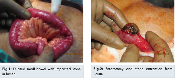
The first patient was 77 year old male. He came to us with classical symptoms of intestinal obstruction since 3 days. He presented with generalized colicky abdominal pain, persistent projectile bilious vomiting, constipation and abdominal distension. On presentation our patient was dehydrated with tachycardia and tachypnea. Abdomen examination showed nontender abdomen with generalized distension with tympanic note. He had past history of ischemic heart disease and was operated for right hydrocele. He was diagnosed with gallstones 2 years back but was not operated by doctors due to poor general and cardiac condition. On admission, routine investigation including liver function test and amylase were normal. Ultrasound was suggestive of dilated bowel loops with pneumobilia and sludge in gall bladder. C.T. scan showed presence of two large 5x4 cm and 4x3 cm gall stones in distal ileum causing complete obstruction of intestines. Gallbladder was empty. There was air in biliary tree with cholecystoduodenal fistula. Hence a diagnosis of intestinal obstruction due to gall stones was presumed. The patient was posted for surgery after correcting electrolyte abnormalities and dehydration. Diagnostic laparoscopy confirmed findings of CT scan, an impacted stone in distal ileum [Fig.1]. Obstructed segment of ileum was delivered through mini-laparotomy. Stones were delivered through enterotomy and enterotomy was subsequently closed [Fig.2]. No further procedure for gallbladder was carried out in view of unstable general condition.
The second patient was 55 year old female who presented with central colicky abdominal pain, copious bilious vomiting, constipation and abdominal distension since 5 days. Her past history was noncontributory. On presentation, she was dehydrated due to persistent vomiting, with tachycardia and hypotension. Per abdomen examination showed mild generalized tenderness with tympanic note on percussion. Clinical impression of acute intestinal obstruction was made. But outside sonography was suggestive of cholelithiasis. In view of contradictory findings on ultrasound, a C.T. scan was carried out. It showed features of small bowel obstruction with a large gall stone in distal ileum measuring 5x3 cm. There was air in biliary tree and sludge in gall bladder with cholecystoduodenal fistula. After stabilization patient was posted for surgery. Initial diagnostic laparoscopy showed small bowel obstruction with a impacted stone in distal ileum. The site of cholecystoenteric fistula was densely covered with adhesions of omentum. A 5 cm mini-laparotomy along with enterotomy was carried out to remove impacted stone from ileum and enterotomy was closed with 3-0 mersilk suture. Post-operative recovery was uneventful. Further procedure was not carried out due to dense adhesions at cholecystoduodenal fistula.
Discussion
Both of our patients were elderly, had stone of large size > 4 cms. Since it was emergency surgery further attempts for gallbladder surgery were avoided. In both cases, gall bladder was covered with omentum with dense adhesion at Calot triangle. Both had classical symptoms of acute intestinal obstruction. Initial laparoscopy was carried to localize the site of obstruction. Through small 5 cm laparotomy incision loop was brought out and enterotomy was carried out. It saved patients of big scar, pain and delayed recovery. Till now patients have remained symptom free.
GI is more common in women, and the ratio of females to males is 3.5 to 1 [
2]. The gallstone must be = 2-2.5 cm in diameter to cause obstruction. As shown by Reisner and Cohen, impaction of the stone can occur in any part of the bowel, i.e., the ileum (60.5% of cases), jejunum (16.1%), stomach (14.2%), colon (4.1%), and duodenum (3.5%). It can also be passed spontaneously (1.3%) [
3,
4]. It occurs most frequently in the terminal ileum [
4].
Classical findings on plain abdominal radiography include: (i) pneumobilia; (ii) intestinal obstruction; (iii) an aberrantly located gallstone; and (iv) change of location of a previously observed stone. In the past, confirming the diagnosis was difficult, but the advent of CT and magnetic resonance imaging (MRI) has made it easier to diagnose GI [
4,
5]. Management of GI is controversial and includes: (i) Enterotomy with stone extraction alone; (ii) Enterotomy, stone extraction, cholecystectomy and fistula closure; (iii) bowel resection alone; and (iv) bowel resection with fistula closure [
2,
5].
Enterotomy with stone extraction alone remains the most common surgical method because of its low incidence of complications [
2]. Spontaneous closure of the fistulous tract is observed in > 50% of cases [
1]. However, 5% of patients who undergo enterolithotomy alone go on to develop biliary symptoms, and 10% require an unplanned reoperation. In the presence of residual stones, the estimated prevalence of recurrence ranged from 5% to 17%, and more than half of these recurrences occur within 6 months of the index presentation. Retrospective cohort and literature reviews of GI reveal a prevalence of biliary malignancy of 2%-6% [
2]. Laparoscopy-assisted methods have been reported by Sarli et al. [
6], who successfully treated three women with GI. Their patients made uneventful recoveries [
6].
If the gallbladder is preserved at the initial procedure, delayed cholecystectomy must be addressed. This is because 5% of patients who have undergone enterolithotomy alone go on to develop biliary symptoms, and the risk of patent fistula reflux and resulting biliary malignancy [
2,
3].
Conclusion
GI is a rare condition affecting mainly the older population. The advent of computed tomography and magnetic resonance imaging has made it easier to diagnose GI. Enterotomy with stone extraction alone remains the most common surgical method because of its low incidence of complications.
References
- Xin-Zheng Dai, Guo-Qiang Li, Feng Zhang, Xue-Hao Wang, Chuan-Yong Zhang. Gallstone ileus: Case report and literature review. World J Gastroenterol. 2013;19(33):5586-5589.
- Halabi WJ, Kang CY, Ketana N, Lafaro KJ, Nguyen VQ, Stamos MJ, et al. Surgery for Gallstone Ileus: A Nationwide Comparison of Trends and Outcomes. Ann Surg. 2014;259(2):329-335
- Reisner RM, Cohen JR. Gallstone ileus: a review of 1001 reported cases. Am Surg. 1994;60:441-446.
- Gupta M, Goyal S, Singal R, Goyal R, Goyal SL, Mittal A. Gallstone ileus and jejunal perforation along with gangrenous bowel in a young patient: A case report. N Am J Med Sci. 2010;2:442-443.
- Ripollés T, Miguel-Dasit A, Errando J, Morote V, Gómez-Abril SA, Richart J. Gallstone ileus: increased diagnostic sensitivity by combining plain film and ultrasound. Abdom Imaging. 2001;26:401-405.
- Sarli L, Pietra N, Costi R, Gobbi S. Gallstone ileus: Laparoscopic- assisted enterolithotomy. J Am Coll Surg. 1998;186:370-371.