Introduction
Ingestion of foreign bodies can be found in all age groups but more common in children, edentulous, alcoholics and psychiatric patients [
1]. Next to nose and throat, oesophagus is the most common site for foreign body impaction [
2]. The commonly ingested foreign bodies include: coins, toy parts, jewellery, batteries, fish or chicken bones and sharp materials such as needles and pins [
1,
3]. The incidence of toothbrush ingestion is however low worldwide with no documented case of spontaneous passage [
3]. A plethora of symptoms and complications are associated with foreign body ingestion. These complications include: perforation, gastrointestinal bleeding or obstructions but some cases are asymptomatic [
4].
Case Report
A 6 year old boy was referred to our facility on account of foreign body ingestion. He was said to have swallowed the whole length of an adult toothbrush whilst mother was brushing his teeth in the morning of presentation to the centre. Despite this, he ate his breakfast of tea with bread without pains.
On examination, he was in stable condition; not in any distress, a temperature of 37.30C, anicteric and acyanosed. There was no pallor or peripheral lymphadenopathy. His respiratory rate was 22/min and the chest was clinically clear. His pulse rate was 94 bpm, normal volume and regular with heart sounds I and II only heard. Abdomen was full, moved with respiration and soft. There was mild tenderness in the right upper abdomen with a firm slender mass palpated in that region. He had no organomegaly and bowel sounds were normoactive. The other systems were essentially normal.
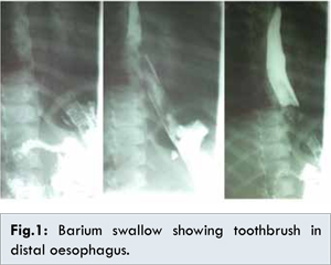
His laboratory test result was as follows: packed cell volume 32%, serum electrolytes: sodium 134.2 mmol/L, potassium 5.4 mmol/L, chloride 114.2 mmol/L, total calcium 2.34 µmol/L, blood urea 4.5 mmol/L, and blood pH 7.5. A plain abdominal X-ray done revealed no radiopaque foreign body, however barium swallow revealed the whole length of a tooth brush in the distal oesophagus with the brush component in the fundus of the stomach [Fig.1].
Child was booked for emergency endoscopy and placed on nil per oral regime. An intravenous line was set and 4.3% dextrose saline infusion was administered. Antibiotic prophylaxis of intravenous ceftriaxone one gram stat was given. Under general anaesthesia using propofol and endotracheal intubation, foreign body was removed using a snare loop through a gastroscope (Olympus Japan) [Fig.2]. He was observed for 2 hours and discharged home on diclofenac suppositories same day when fully conscious. An instruction of nil per oral till a gastrografin swallow next morning was given. He was seen in the morning of 1st day post-procedure in very stable condition. Parents declined further radiological test due to financial constraint. A graded oral sip of water was cautiously commenced. His management was continued on out-patient basis with graduation from fluid to solid diet with no complaint. A 7th day follow up visit was uneventful.
Discussion
Majority of foreign body ingestion occurs in the paediatric population with a peak age of 6 months to 6 years [
5]. Perforation, obstruction or impactions usually occur at anatomical narrowing in the gastrointestinal tract. At least a third of the foreign bodies retained in the gastro-intestinal tract are present in the oesophagus [
6]. Once through the oesophagus 80-90% of foreign bodies pass out uneventfully but 10-20% require endoscopy and <1% require surgery [
7,
8]. Foreign bodies longer than 10 cm such as the tooth brush cannot negotiate the c-loop of the duodenum successfully due to its fixed retroperitoneal position [
7]. A prompt diagnosis is usually based on the history, physical examination and radiological investigations.
In Port Harcourt Nigeria, studies revealed the common foreign bodies ingested as dentures, metallic objects and fish bone with no case of tooth brush ingestion [
9,
10] while in Enugu Nigeria [
11] coin is the most common. Globally, the most common ingested foreign body in the paediatric population is the coin [
1,
7,
8].
The clinical features can be varied depending on time to presentation, age group, type and location of the foreign body. Frequently, symptoms occur well after the patient ingests the foreign body. These symptoms may include choking, refusal to eat, vomiting, drooling, wheezing, blood-stained saliva, or respiratory distress [
12]. Oropharyngeal or proximal oesophageal perforation can cause neck swelling, erythema, tenderness, or crepitus [
12]. 75% of children have impacted foreign bodies located at the upper oesophageal sphincter while 75% of adults have it located at the lower oesophageal sphincter [
11]. Of significance in this report was the absence of food impaction despite the size of foreign body relative to the paediatric oesophageal lumen.
Radiological investigations are of immense value as they can detect the location, size, shape and number of foreign bodies. The conventional radiographs demonstrate radiopaque foreign bodies. Studies have shown that 60%-65% of foreign bodies are radiopaque [
13]. Limitations of radiological investigation include non-visualization of radiolucent objects, radiation exposure and failure of precise location during removal. Views taken include a soft tissue neck in cases where impaction at the neck is suspected, a posterior-anterior and lateral chest radiograph and if equivocal an abdominal radiograph. The toothbrush swallowed was radiolucent.
Barium or gastrografin swallow are indicated in cases of radiolucent foreign body ingestion as was observed in this case report. These contrast studies can obscure future endoscopy site, increase risk of aspiration and is contraindicated if there is suspected perforation thus are not routinely done. Gastrographin is preferred where oesophageal perforation is suspected. This is because sipping of barium sulphate into the mediastinum in perforation remains there and can cause granuloma while gastrographin is absorbed in the circulatory system. Contrast studies are more valuable post-removal of foreign body to rule out strictures and pseudodiverticulum [
13]. Ultrasonography is a first line choice for foreign bodies in soft tissues but limited if the foreign body is in the gastrointestinal tract due to bowel gas that can obscure the object [
14]. A computerized tomography scan also plays a role in radiolucent foreign bodies that are not detected on radiographs [
12].
Once diagnosis has been made, management decisions depend on several factors like the age of the patient, clinical condition, anatomical location, size/shape of foreign body and what interventional procedures are readily available [
15]. Oesophageal foreign bodies require prompt removal within 24 hours because delay increases the likelihood of injuries like strictures, trachea-oesophageal and aorto-esophageal fistulas when the object erodes the mucosa. This in turn decreases the chances of successful removal [
3,
7]. There has been no record of spontaneous passage of toothbrush till date. Other indications for removal of foreign bodies include symptomatic patients, sharp or long objects > 5 cm (as in the index case), magnet, disc battery and if object is in the stomach > 4-6 weeks [
8,
15].Technique of removal will depend on the location/ type of foreign, patient’s condition and surgeon’s expertise. The different techniques that could be employed include flexible or rigid endoscopy, Magill forceps and laryngoscope, Foley’s catheter, bougienage, penny pincher, laparoscopy and open surgery.
Flexible endoscopy has been seen to be the most successful method and allows visualization of sharps. Studies have reported successful endoscopic removal of tooth brushes and other objects [16-19]. A successful endoscopic retrieval in this report was recorded using a snare loop as an endoscope accessory [Fig.2]. Chiu et al. in a study done in Taiwan had a success rate of 96.4%. Rat tooth forceps and snares were the most frequently used flexible endoscope accessories in their study. In cases of unsuccessful removal by endoscopy, surgical techniques should be sought. Jamal et al. [20] reported a case of successful laparoscopic removal of a brush from the stomach. Complications of endoscopic retrieval include sore throat, lacerations, perforation, oesophageal stricture and diverticula. Perioesophageal abscess, mediastinitis, pneumothorax, pleural effusion and respiratory arrest are other complications. In the index case only a transient sore throat was recorded. Most death from foreign body perforation of the gastrointestinal tract is due to fulminant sepsis. There is excellent prognosis if foreign bodies are removed promptly but increased morbidity and mortality with chronic cases.
Conclusion
Ingested toothbrush cannot pass spontaneously through the gastrointestinal tract therefore prompt removal is of essence to avoid complications. Flexible endoscopy is very useful in the retrieval of ingested foreign body in children.
References
- Ikenberry SO, Jue TL, Anderson MA, Appalaneni V, Banerjee S, Ben-Menachem T, et al. Management of ingested foreign bodies and food impactions. Gastrointest Endosc. 2011;73(6):1085-1091.
- Akhtar M, Hag MI. Management of oesophageal foreign bodies. Professional Med J. 2008;12(13):308-331.
- Faust J, Schreiner O. A swallowed toothbrush. Lancet. 2001;357:1012.
- Sung SH, Jeon SW, Sun HS. Factors predictive of risk for complication in patients with esophageal foreign body. Dig Liver Dis. 2011;43(8):632-635.
- Webb WA. Management of foreign bodies of the upper gastrointestinal tract: update. Gastrointest Endosc. 1995;41:39-51.
- Muhammad R, Khan Z, Jamil A, et al. Frequency of oesophageal foreign bodies and their sites of impaction in patients presenting with foreign body in aerodigestive tract. European Scientific Journal. 2013;9(21):218-220.
- Li ZS, Sun ZX, Zou DW, Xu GM, Wu RP, Liao Z. Endoscopic management of foreign bodies in the upper-GI tract: Experience with 1,088 cases in China. Gastrointest Endosc. 2006;64:485-489.
- Eisen GM, Baron TH, Dominitz JA, Faigel DO, Goldstein JL, Johanson JF, et al. Guideline for the management of ingested foreign bodies. Gastrointest Endosc. 2002;55:802-806.
- Nwogbo AC, Eke N. Oesophageal foreign bodies in Port Harcourt. Port Harcourt Med J. 2012;6(2):211-214.
- Ibekwe MU, Onotai LO, Otaigbe B. Foreign body in the ear, nose and throat in children: A five year review in Niger Delta. Afr J Paediatr Surg. 2012;9(1):3-7.
- Oriji FT, Akpeh JO, Okolugbo NE. Management of Esophageal Foreign Bodies: Experience in a Developing CountryWorld J Surg. 2012; 36:1083-1088.
- Conway WC, Sugawa C, Ono H, Lucas CE. Upper GI foreign body: an adult urban emergency hospital experience. Surg Endosc. 2007;21:455-460.
- Lee JH, Kim HC, Yang DM, Kim SW, Jin W, Park SJ, Kim HJ. What is the role of plain radiography in patients with foreign bodies in the gastrointestinal tract? Clin Imaging. 2012;36:447-454.
- Piotto L, Gent R, Kirby CP, Morris LL. Preoperative use of ultrasonography to localize an ingested foreign body. Pediatr Radiol. 2009;39:299-301.
- Harvey M, Cave G, Prince G. Endoscopic removal of an inadvertently swallowed toothbrush in the emergency department. Case Rep Emerg Med. 2012;2012:568163.
- Chiu YH, Hour SK, Chen SC, How CK, Lam C, Kao WF, Yen DH, Huang MS. Diagnosis and endoscopic management of upper gastrointestinal foreign bodies. Am J Med Sci. 2012;3(43):192-195.
- Lu XL, Cao HL, Qian KD. Endoscopic removal of an accidentally swallowed toothbrush. Intern Med. 2008;47:1797-1798.
- Niknam R, Mahmoudi L, Nasseri -Moghaddam S. Swallowed Tooth Brush. Arch Iran Med. 2012;15(3):177-178.
- Xu L, Huang C, Qu C, Zhang Y, Zhou M, ChenY. Easy and effective endoscopic retrieval of ingested sharp foreign bodies. J Basic Appl Sci. 2013;9:87-90.
- Jamal K, Shaunak S, Kalsi S, Nehra D. Successful Laparoscopic Removal of an ingested toothbrush. Journal of Surgical Technique and Case Report. 2013;5(2):99-102.