|
|
|
|
|
Well Differentiated Neuroendocrine Carcinoma of the Testis in a Nigerian Male
|
|
|
Nzechukwu Zimudo Ikeri1, Feziechukwu Collins Nnaji2, Olayemi Dawodu1, Uche Igbokwe3, Fatimah Biade Abdulkareem1, Adekunbiola Aina Banjo1
1Department of Anatomic and Molecular Pathology, Lagos University Teaching Hospital, Nigeria, PMB 12003, Lagos, Nigeria; 2Simeon Hospital, 83 Sanya Street, Aguda, Surulere Lagos; 3Queen’s Hospital, Romford, Essex, United Kingdom. |
|
|
|
|
|
Corresponding Author:
|
Dr. Nzechukwu Zimudo Ikeri
Email: zimudo@gmail.com
|
|
|
|
|
|
|
|
|
Received:
08-JUL-2015 |
Accepted:
09-OCT-2015 |
Published Online:
05-NOV-2015 |
|
|
|
|
|
|
|
Abstract
|
|
|
|
Testicular carcinoids are very rare tumors that account for less than 1% of all testicular neoplasms. We present the first case of a testicular carcinoid tumor in a Nigerian male who had radical orchiectomy on account of a painful left testicular mass. Histology showed a thinly encapsulated lesion composed of sheets of closely packed spindle cells having a uniform ‘salt-and-pepper’ chromatin pattern, and moderate pale cytoplasm within a scant dense fibrocollagenous stroma. Mitoses were scanty and necrosis absent. There was no evidence of an intratubular germ cell tumor. The lesional cells stained positive, on immunohistochemistry, for CD 56, NSE and S100, though they were negative for chromogranin A and synaptophysin. They also showed positivity for AE 1/3 in a para-nuclear dot-like fashion. There was complete negativity for CD 99, CD 117, PLAP and EMA. The cells were weakly positivity for inhibin, while CD 45 marked only a small population of lymphocytes. A diagnosis of a spindle cell type carcinoid tumour/well differentiated neuroendocrine carcinoma was made. The presence of a fibrous capsule suggested a primary testicular lesion and subsequent abdomino-pelvic CT scan showed no intra-abdominal primaries. However, investigations to rule out a focus from the lungs are yet to be done. |
|
|
|
|
|
Keywords :
|
Carcinoid Tumor, Neuroendocrine Carcinoma, Germ Cell Neoplasms, Orchiectomy, Testicular Neoplasms.
|
|
|
|
|
|
|
|
|
|
|
|
6go6ckt5b8|3000F7576AC3|Tab_Articles|Fulltext|0xf1ffa4a1090000003104000001000500 6go6ckt5b5idvals|533 6go6ckt5b5|2000F757Tab_Articles|Fulltext Introduction
Testicular neuroendocrine tumors are very rare accounting for 0.23% of all testicular neoplasms [ 1]. They most commonly have their origin within the testis either in pure form or in association with a germ cell tumor, but could also be metastases from the gastrointestinal tract or lungs [ 2]. Testicular carcinoids most often present as a painless testicular mass or enlargement; only a few cases present with carcinoid syndrome [ 1, 3]. Like carcinoid tumors in other sites, they could have typical or atypical histopathology, which can predict their clinical behaviour [ 3]. We present the first case of testicular carcinoid tumor in a Nigerian male who had radical orchiectomy on account of a painful left testicular mass. Histology showed spindle cell type carcinoid tumor which was confirmed by immunohistochemistry.
Case Report
A 35 year old man presented to the outpatient clinic of a private hospital with a one month history of pain in the left testis. There was no prior history of trauma. On physical examination, a tender left testicular mass was felt. Ultrasonography of the testes revealed an enlarged left testicle that measured 53x30 mm, with a homogenous hypoechoic vascular mass located in its superior pole. The mass measured 38x25 mm. Spectral broadening was noted on pulse Doppler ultrasonography. The right testis was unremarkable. A presumptive diagnosis of germ cell tumor probably a seminoma was made and total left orchiectomy was performed.
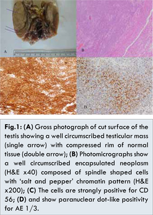
Gross examination of the testicular specimen revealed a well circumscribed mass measuring 4x2.5x2 cm with a rim of compressed normal testicular tissue [Fig.1a]. Histopathologic assessment revealed a thinly encapsulated lesion composed of sheets of closely packed spindle cells with scant dense fibrocollagenous tissue stroma. The lesional cells were markedly uniform with ‘salt and pepper’ chromatin pattern and moderate amount of pale cytoplasm [Fig.1b]. Mitoses were scanty (1 per 10 high power fields) and there was no necrosis. The adjacent seminiferous tubules displayed spermatogenesis with no evidence of intatubular germ cell neoplasm. A preliminary diagnosis of spindle cell neoplasm probably malignant was entertained.
Immunohistochemistry showed complete negativity for CD 117 and PLAP, thus excluding a germ cell tumor. CD45 marked only a very small population of lymphocytes. There was only a weak patchy expression of inhibin which with the absence of longitudinal nuclear grooves and Call-Exner bodies ruled out an adult granulosa cell tumor. A possibility of Sertoli cell tumor was entertained (including its sarcomatoid form), however the absence of any structures resembling tubules and complete negativity with CD99 excluded this. The cells were also negative for TTF-1, calcitonin and Melan A. Though the cells were negative for chromogranin A and synaptophysin, they showed strong uniform expression for CD56 [Fig.1c] and a moderate patchy expression of NSE and S100. They also showed positivity for broad spectrum cytokeratins (AE 1/3) in a para-nuclear dot-like fashion [Fig.1d]. These immunohistochemical findings when taken together with the morphological characteristics were considered to be strongly indicative of a spindle cell type carcinoid tumor/well differentiated neuroendocrine carcinoma. The presence of a fibrous capsule indicates that the tumor was most likely a primary, but the patient was advised to undergo more investigations to exclude a primary source elsewhere. The patient has undergone an abdominopelvic CT scan which revealed no tumor deposits in the gastrointestinal tract, liver or other intra-abdominal organs. He is yet to undergo investigations to exclude a primary from the lungs.
Neuroendocrine tumors of the testis are very rare, accounting for less than 1% of testicular neoplasms [ 2]. As a result, limited studies have been published and none as yet has been documented in the Nigerian population. They consist of three groups of tumors: primary testicular carcinoid, carcinoid differentiation within a germ cell tumor (usually a mature cystic teratoma) and metastatic carcinoid from extra-testicular sources, most commonly the gastrointestinal tract [ 1, 2]. Primary testicular carcinoid, as was seen in the index patient, is the commonest of these three categories. Stroosma et al. in their review of 62 cases, reported 44 (71%) to be primary, 6 (9.7%) to be metastases from other locations and 12 (19.4%) to arise within a teratoma [ 4]. In another study of 10 cases, all were found to be of testicular origin [ 5]. There was no teratomatous component in the index case presented. Carcinoids have also been reported to occur in cryptorchid testes as well as in a case with ovotestis [ 6- 8]. Due to lack of morphologic differences between primary and metastatic carcinoids, it is necessary to exclude the presence of primary disease elsewhere before making a diagnosis of primary testicular carcinoid by employing the use of barium contrast studies, computerized tomography, somatostatin receptor scintigraphy etc [ 1, 2].
Testicular carcinoids have been reported to occur in a wide age group ranging from 10 to 83 years [ 2]. The mean age group however is 40-60 years [ 9]. Primary testicular carcinoids were once thought to arise from Argentaffin cells within the gonad, but this notion has been discarded due to the absence of these cells in the testis [ 10]. Some workers have proposed the origin of primary testicular carcinoids from Leydig cells, however Abbosh et al. showed identical genetic aberrations within primary testicular carcinoids and coexisting mature teratoma [ 11, 12]. Primary testicular carcinoid tumors are now thought to be of germ cell origin, arising either as a monodermal germ cell tumor or existing as the remaining component of a burnt out teratoma [ 2].
Testicular carcinoid presents most commonly as a painless unilateral mass/swelling of the testis [ 1]. It affects the left testis more commonly than the right, as was the case in the index patient. There can however be bilateral testicular involvement or associated pain [ 13, 14]. One unusual case was reported with a mass in one testis and pain in the contralateral testis [ 15]. The index case presented with a painful left testicular mass. Few patients are asymptomatic, and fewer still present with carcinoid syndrome. In one study, 62.5% of cases presented as a mass, 25% were discovered incidentally while 8.3% presented with carcinoid syndrome [ 3]. In the study conducted by Reyes et al. 6 of 10 patients presented with a painless mass, while the rest presented with tender swelling. There was no asymptomatic case or any that presented with carcinoid syndrome [ 5]. In another study of 7 cases, all presented with painless testicular masses and none had carcinoid syndrome [ 9]. Carcinoid syndrome when present is due to bioactive substances secreted into the blood stream resulting in diarrhea, skin flushes, asthma and palpitations [ 2, 16].
Carcinoids though mostly benign in behavior have a malignant potential, and cases of metastases have been documented. Cases can therefore be staged according to the American Joint Committee on Cancer (AJCC) criteria [ 5]. Metastases however are rare in primary testicular carcinoids. Stroosma reported metastases in 15.9% of cases of primary testicular lesions [ 4]. Wang et al. reported metastases in none of 20 typical primary testicular cases and in 1 of 4 cases with atypical morphology [ 3]. Documented sites of metastases include the para-aortic lymph nodes, lungs, vertebrae, retroperitoneum, skin and skeletal muscle [ 17- 20].
Grossly, testicular carcinoids have been reported to measure between 1 and 9.5 cm with a mean size of 4.6 cm. They are solid and lobulated, yellow to dark-tan and may have calcifications [ 20]. Histologically, testicular carcinoids show a predominance of trabecular and insular architecture; and large tumors may show necrosis [ 20]. In grade 1 tumors (G1), cells are monomorphic with round nuclei, finely dispersed chromatin, acidophilic finely granular cytoplasm, low mitotic activity (<2/10 high power fields) and no necrosis [ 21, 22]. Higher grades feature more mitoses, necrosis, and cellular pleomorphism. Grading of testicular carcinoid correlates well with the clinical outcome and metastases are seen more commonly in atypical cases [ 22]. Histologic assessment of our case, here presented, revealed no atypical features.
Testicular carcinoids show positive immunostaining for neuroendocrine markers like chromogranin, synaptophysin, NSE and cytokeratins (CK) [ 9, 23]. However they are not always positive for these markers [ 24, 25]. Carcinoids also stain strongly positive for CD 56, which was so for our case presented [ 26]. Though carcinoids are presumed to be of germ cell origin, they do not show significant numerical abnormalities of the X chromosome as in other testicular germ cell tumors [ 27]. Cytogenetic abnormalities reported in testicular carcinoids include aneuploidy, tetraploidy, isochromosome 12 and 12p over-expression [ 12, 28].
Treatment of localized testicular carcinoid is primarily surgical, either by focal excision or radical orchiectomy [ 3]. Chemotherapy and adjuvant radiotherapy have been employed in the treatment of metastatic cases [ 3]. Most carcinoids are benign with favourable prognosis. However, long-term prognosis is dependent on histologic grade. Wang et al. in their study showed that after a mean follow up of 52.7 months, all cases with typical histology were alive without recurrence or metastases, while 25% of those with atypical histology showed metastases [ 3]. Increasing size, association with teratoma and metastases are also poor prognostic factors. Metastases have been reported to occur as long as 6 years after orchiectomy, indicating the need for long term follow up with urinary 5-hyddroxyindoleacetic acid (5-HIAA) or serum chromogranin A [ 29].
Conclusion
Testicular carcinoids are very rare and this is the first reported case in a Nigerian patient. The presence of primary disease elsewhere must always be excluded before making a diagnosis of primary testicular carcinoid. Long-term follow up is essential for detecting recurrence or metastatic disease.
Acknowledgement
The authors would like to acknowledge Mr Adeteye, Olawale Victor for taking the photographs of the specimen.
References
- Chang YH, Chuang CK, Wu CT, Ng KF, Liao SK. Primary carcinoid tumor of the testis: case report. Chuang Gung Med J. 2002;25(10):695-699.
-
Neely D, Gray S. Primary Carcinoid tumour of the testis. Ulster Med J. 2011;80(2):79-81.
-
Wang WP, Guo C, Berney DM, Ulbright TM, Hansel DE, Shen R, et al. Primary carcinoid tumours of the testis: a clinicopathologic study of 29 cases. Am J Surg Pathol. 2010;34(4): 519-524.
-
Stroosma OB, Delaere KP. Carcinoid tumours of the testis. BJU Int. 2008;101(9):1101-1105.
-
Reyes A, Moran CA, Suster S, Mchal M, Dominguez H. Neuroendocrine carcinomas (carcinoid tumour) of the testis. A clinicopathologic and immunohistochemical study of ten cases. Am J Clin Pathol. 2003;120(2):182-187.
-
Kim JH, Noh TI, Shim JS, Ham BK, Choi H, Bee JH, et al. Primary testicular carcinoid tumour with mature teratoma in undescended testis metastatic to lymph nodes. Can Urol Assoc J. 2014(3-4):E245-248.
-
Finci R, Gunhan O, Celasun B, Gungor S. Carcinoid tumor of undescended testis. J Urol. 1987;137(2):301-302.
-
Esen T, Erdogru T, Muslumanoglu M, Kilicaslan I, Kayserili H. Laparoscopic removal of the uterus, seminal vesicle and bilateral ovotestes harbouring mature teratoma and carcinoid tumor in an intersex patient. J Urol. 1996;155(6):2032-2033.
-
Liu FF, Zheng JF, Zhou LT, Wang CC, Wang JJ, Shen Q, et al. Primary neuroendocrine tumour of the testis: clinicopathological study of 7 cases. Zhonghua Nan Ke Xue. 2014;20(1):63-67.
-
Merino J, Zuluaga A, Gutierrez-Tejero F, Del Mar Serrano M, Ciani S, Nogales FF. Pure testicular carcinoid associated with intratubular germ cell neoplasia. J Clin Pathol. 2005;58(12):1331-1333.
-
Mai KT, Park PC, Yazdi HM, Carlier M. Leydig cell origin of testicular carcinoid tumour: immunohistochemical and electron microscopic evidence. Histopathology. 2006;49(5):548-549.
-
Abbosh PH, Zhang S, Maclennan GT, Montironi R, Lopez-Beltran A, Rank JP, et al. Germ cell origin of testicular carcinoid tumors. Clin Cancer Res. 2008;14(5):1393-1396.
-
Son HY, Ra SW, Jeong JO, Koh EH, Lee HI, Koh JM, et al. Primary carcinoid tumor of bilateral testis associated with carcinoid syndrome. Int J Urol,. 2004;11(11):1041-1043.
-
Saxena A, Watkin SW. Bilateral malignant testicular carcinoid. Br J Urol. 1990;65(3):302-303.
-
Palla AR, Hogan T, Singh S. Unusual presentation of a left testicular carcinoid. Case Rep Oncol. 2012;5(1):43-46.
-
Han X, Yu L, Yang S, Zheng J. Primary neuroendocrine tumour of the testis: a study of clinicopathologic features. Int J Clin Exp Pathol. 2014;7(4):1771-1776.
-
Pectasides D, Glotsos J, Bountouroglou NG, Dadioti PA, Athanassiou AE. Primary carcinoid of the testis with metastases. Case report and review of literature. J BUON. 2002;7(2):153-156.
-
Abrahamsson J, Mellander L, Nilsson O, Rubensson A. Multiple lymph node metastases in a boy with primary testicular carcinoid, despite negative preoperative imaging procedures. J Pediatr Surg. 2005;40(11):e19-21.
-
Shimura S, Uchida T, Shitara T, Nishimura K, Murayama M, Honda N, et al. [Primary carcinoid tumour of the testis with metastases to the upper vertebrae. Report of a case]. Nihon Hinyokika Gakkai Zasshi. 1991;82(7):1157-1160.
-
Mostofi FK, Sesterhenn IA, Srigley JR, Levin HS. Tumours of the testis and paratesticular tissue. In: Ebele JN, Sauter G, Epstein JI, Sesterhenn IA (eds). Pathology and genetics of tumours of the urinary system and male genital organs. IARC press, Lyon. 2004: pp. 217-278.
-
Tumours of the Male genital tract, In: Fletcher CD (ed). Diagnostic histopathology of tumours pp. 733-838. 2nd edition, Churchill Livingston.
-
Strosberg JR, Nasir A, Hodul P, Kvols L. Biology and treatment of Metastatic Gastrointestinal Neuroendocrine Tumours. Gastrointest Cancer Res. 2008;2(3):113-125.
-
Zhoa YC, Shi QL, Zhao XJ, Ma HH, Lu ZF, Zhou HB. [Clinicopathologic study of primary carcinoid tumour of the testis]. Zhonghua Nan Ke Xue. 2007;13(2):157-160.
- Jensen SM, Gazdar AF, Cuttitta F, Russell EK, Linnoila RI. A comparison of synaptophysin, chromogranin, and L-decarboxylase as markers for neuroendocrine differentiation in lung cancer cell lines. Cancer Res. 1990;50(18):6068-6074.
-
Terada T. Carcinoid tumours of digestive organs: a clinicopathologic study of 13 cases. Gastroenterol Res. 2009;2(1):35-37.
-
Special techniques in surgical pathology. In: Houston M (ed) Rosai and Ackerman’s surgical pathology pp. 37-94. 10th edition. Elsevier Mosby, Edinburg.
-
Kato N, Motoyama T, Kameda N, Hiruta N, Emura I, Hasegawa G, et al. Primary carcinoid tumour of the testis: Immunohistochemical, ultrastructural and FISH analysis with review of the literature. Pathol Int. 2003;53(10):680-685.
-
Zavala-Pompa A, Ro JY, el-Naggar A, Ordonez NG, Amin MB, Pierce PD, et al. Primary carcinoid tumour of testis. Immunohistochemical, ultrastructural, and DNA flow cytometric study of three cases with a review of the literature. Cancer. 1993;72(5):1726-1732.
-
Sasaki M, Emura M, Kim U, Shinbo M, Shima T, Suzuki N, et al. Primary carcinoid tumour of the testis metastatic to the para-aortic lymph nodes in six years after the first operation: a case report. Hinyokika Kiyo. 2009;33(4):233-236.
|
|
|
|
|
|
|
Search Google Scholar for
|
|
|
Article Statistics |
|
Ikeri NZ, Nnaji FC, Dawodu O, Igbokwe U, Abdulkareem FB, Banjo AAWell Differentiated Neuroendocrine Carcinoma of the Testis in a Nigerian Male.JCR 2015;5:473-478 |
|
Ikeri NZ, Nnaji FC, Dawodu O, Igbokwe U, Abdulkareem FB, Banjo AAWell Differentiated Neuroendocrine Carcinoma of the Testis in a Nigerian Male.JCR [serial online] 2015[cited 2026 Jul 22];5:473-478. Available from: https://www.casereports.in/articles/5/2/Well-Differentiated-Neuroendocrine-Carcinoma-of-the-Testis-in-a-Nigerian-Male.html |

|
|
|
|
|