|
|
|
|
|
Naso-Ophthalmic Myiasis and Pubic Louse Infestation of Nose
|
|
|
Kavitha Duraipandi1, Nitin Gupta2, Anjana Karunakaran1, Pratibha Kale2, Sabin Dhaka1, Nishant Verma2, Bijay Ranjan Mirdha2
Departments of Ophthalmology1 and Microbiology2, All India Institute of Medical Sciences, New Delhi- 110029, India. |
|
|
|
|
|
Corresponding Author:
|
Dr. BR Mirdha
Email: mirdhabr2078@gmail.com
|
|
|
|
|
|
|
|
|
Received:
21-OCT-2015 |
Accepted:
07-DEC-2015 |
Published Online:
05-JAN-2016 |
|
|
|
|
|
|
|
Abstract
|
|
|
|
A 15 year old girl presented to the emergency with complaints of foreign body sensation in eyes. Multiple translucent larvae were observed on the conjunctivae. A nasal cavity search also revealed similar larvae localised near the opening of nasolacrimal duct in the inferior meatus. They were identified as Oestrus ovis, which can affect humans causing cavitary myiasis, affecting eyes and rarely nose. In addition, presence of Pthirus pubis was also found in the nasal cavity. Pthirus pubis infestation has been reported from pubic hairs, axillary hairs, eyelashes, moustache and beard but never from the nasal cavity. This is a very rare case of concomitant ophthalmic and nasal myiasis along with pubic louse infestation of nasal cavity. |
|
|
|
|
|
Keywords :
|
Foreign Bodies, Larva, Myiasis, Nasal Cavity, Nasolacrimal Duct.
|
|
|
|
|
|
|
|
|
|
|
|
6go6ckt5b8|3000F7576AC3|Tab_Articles|Fulltext|0xf1ff24da0a0000005a04000001000c00 6go6ckt5b5idvals|557 6go6ckt5b5|2000F757Tab_Articles|Fulltext Introduction
Myiasis is an infestation of humans with dipterous larvae, commonly seen in the developing settings. Cavitary myiasis (e.g nasal and ophthalmic myiasis) is the commonest anatomical type of myiasis [1]. Most cases of human myiasis are caused by flies belonging to following families: Muscidae, Oestridae, Calliphoridae, and Sarcophagidae. Oestrus ovis (Sheep bot fly) belonging to Oestridae family are one of the commonest flies causing myiasis world over. The female fly may deposit hundreds of first-instar larvae at a time, mostly during the summer season. It is known that larvae of O. ovis usually develop in the sinuses and nasal passage of sheep in the sheep farming areas of the world. They occasionally affect humans causing cavitary myiasis, most commonly affecting eyes [2-5]. In humans, larvae of O. ovis do not develop beyond the first instar stage and die within 10 days if not removed. Pthirus pubis infestation of pubic hairs is a common phenomenon in persons living in developing settings and is a clear indicator of poor hygiene and overcrowding. Besides pubic region, adult louse has also been reported from axillary hairs, eyelashes, moustache and beard. Here, we present an unusual case of concomitant ophthalmic and nasal myiasis along with pubic louse infestation of nasal cavity in a young individual.
Case Report
A 15 year old girl presented to the emergency of with complaints of foreign body sensation, watering and redness of left eye along with flushing of the face. However, visual acuity was 6/6 in both the eyes, and pupillary reflexes and ocular movements were normal. On slit lamp examination, lower punctum of the left eye was slightly everted. The conjunctiva was diffusely congested and several translucent larvae were observed on the bulbar conjunctiva, in the lower fornix and palpebral conjunctiva. The cornea, anterior chamber and fundus examination of both the eyes were within normal limits. A saline wash was given to the left conjunctival sac for about 5 minutes.
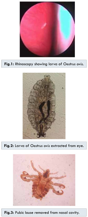
A McPherson’s forceps was used to pick up and remove the larvae from the bulbar conjunctiva, lower palpebral conjunctiva, the medial and lateral canthi. Then the upper lid was everted and larvae were removed. Clockwise screening was done twice from upper fornix, lateral canthus, lower fornix and medial canthus to remove any remaining larvae. A nasal cavity search also revealed similar larvae localised near the opening of nasolacrimal duct in the inferior meatus [Fig.1]. The larvae retrieved from the eye and nasal cavity were examined microscopically. All these had an oval-shaped segmented body with prominent oral hooks connected to the internal cephalopharyngeal skeleton. These larvae were identified as the first instar larva of the sheep botfly, Oestrus ovis [Fig.2]. In addition, presence of an insect was also found in the nasal cavity. The examination of insect under light microscopy revealed wingless, translucent crab like lice with three pairs of legs along with claws. It was confirmed as Pthirus pubis [Fig.3].
Ophthalmomyiasis externa refers to the superficial infestation of ocular tissue where the patient presents with complaints of acute foreign-body sensation with lacrimation whereas nasal myiasis presents as foreign-body sensation, nasal discharge and facial pain [6-8]. Although external ophthalmomyiasis is relatively common in tropical countries, there are only handful of reports of naso-ophthalmic myiasis available in published literature [9,10]. Oestrus ovis, besides other etiological agents has been known to cause nasal myiasis [11,12]. In the present case, it was difficult to establish whether the primary site of infection of O. ovis was nose or eyes. The hypothesis of primary nasal colonisation and secondary infestation of eye with larvae passing through the naso-lacrimal duct could possibly be supported by the everted punctum in the left eye. The other possibility of primary ophthalmic infestation with possible migration through the nasolacrimal duct to the nasal cavity could not be ruled out. Migration of larvae through the lacrimal canal to the nose cavity has been reported previously in published literature [9]. The only definite conclusion that could be drawn was that there was larval migration through the nasolacrimal duct from or to the eyes.
The findings of this case emphasises upon a thorough search of nasal cavity in cases of ophthalmomyiasis and vice-versa. The treatment of myiasis in most cases is mechanical removal of the larvae. In cases of ophthalmomyiasis, if only larvae is removed from the eyes and no search of nasal cavity is done, then nose may serve as the source of larvae for recurrent eye infestations. Pubic louse infestation of the nasal cavity to our knowledge has not been reported in the published literature. It is likely that questionable hygiene and poor socioeconomic condition was linked with the dual infestations.
References
- Zumpt F. 1965. Myiasis in man and animals in the Old World. Butterworths, London, United Kingdom.
- Gregory AR, Schatz S, Laubach H. Ophthalmomyiasis caused by the sheep bot fly Oestrus ovis in northern Iraq. Optum Vis Sci. 2004;81:586-590.
- Jenzeri S, Ammari W, Attia S, Zaouali S, Babba H, Messaoud R, et al. External ophthalmomyiasis manifesting with keratouveitis. Int Ophthalmol. 2008;29:533-535.
- Reingold WJ, Robin JB, Leipa D, Kondra L,Schanzlin DJ, Smith RE. Oestrus ovis ophthalmomyiasis externa. Am J Ophthalmol.1984;97:7-10.
- Sreejith RS, Reddy AK, Ganeshpuri SS, Garg P. Oestrus ovis ophthalmomyiasis with keratitis. Indian J Med Microbiol. 2010;28:399-402.
- González AC, Salamanca GJC, Olano VM, Pérez CE. Cavitary myiasis: case report. Rev Fac Med. 2008;16:95-98.
- Madana J, Yolmo D, Gopalakrishnan S, Saxena SK, Nath AK, Ilamaran V. Hypohidrotic ectodermal dysplasia with atrophic rhinitis and nasal myiasis. Int J Pediatr Otorhinolaryngol. 2009;73:1467-1469.
- Tsang WS, Lee DL. Nasal myiasis: the role of endoscopy. Ear Nose Throat J. 2009;88:1250-1251.
- Eyigor H, Dost T, Dayanir V, Basak S, Eren H. A case of naso-ophthalmic myiasis. Kulak Burun Bogaz Ihtis Derg. 2008;18:371-373.
- Smillie I, Gubbi PK, Cocks HC. Nasal and ophthalmomyiasis: case report. J Laryngol Otol. 2010;124(8):934-935.
- Badia L, Lund VJ. Vile bodies: an endoscopic approach to nasal myiasis. J. Laryngol Otol. 1994;108:1083-1085.
- Lucientes J, Clavel A, Ferrer-Dufol M, Valles H, Peribanez MA, Gracia-Salinas MJ, et al. One case of nasal human myiasis caused by third stage instar larvae of Oestrus ovis. Am J Trop Med Hyg. 1997;56:608-609.
|
|
|
|
|
|
|
Search Google Scholar for
|
|
|
Article Statistics |
|
Duraipandi K, Gupta N, Karunakaran A, Kale P, Dhaka S, Verma N, Mirdha BRNaso-Ophthalmic Myiasis and Pubic Louse Infestation of Nose.JCR 2016;6:1-3 |
|
Duraipandi K, Gupta N, Karunakaran A, Kale P, Dhaka S, Verma N, Mirdha BRNaso-Ophthalmic Myiasis and Pubic Louse Infestation of Nose.JCR [serial online] 2016[cited 2026 May 17];6:1-3. Available from: http://www.casereports.in/articles/6/1/Naso-Ophthalmic-Myiasis-and-Pubic-Louse-Infestation-of-Nose.html |

|
|
|
|
|