6go6ckt5b8|3000F7576AC3|Tab_Articles|Fulltext|0xf1ff6c4b270000004208000001000100
6go6ckt5b5idvals|918
6go6ckt5b5|2000F757Tab_Articles|Fulltext
Introduction
The distal tibiofibular joint is described as a syndesmosis. It comprises the tibia and fibula, the fibrous interosseous membrane between the two bones, the anterior and posterior inferior tibio-fibular ligament, transverse ligament and the interosseous ligament. Syndesmotic ligament injury is relatively uncommon contributing upto 1-18% of ankle ligament injuries [1]. Its incidence significantly increases between
13-50% with associated ankle fracture [2,3]. The huge variation probably is either because of under diagnosis or under reporting [4].
Failure to stabilise syndesmosis results in tibiofibular diastasis. This in consequence leads to lateral displacement and external rotation of talus, thereby decreasing the talotibial contact area and increases the loading pressure. Even 1 mm lateral shift of talus decreases the contact area by 42% [5]. The optimum method of stabilisation of the disrupted syndesmosis remains controversial. Traditionally screw fixation was the treatment of choice. But choice of screw, number of cortices to be engaged, post-operative protocols and removal of implant were debatable.
Alternately syndesmotic tightrope (Arthrex Inc) can be used to stabilise the syndesmosis. This appears to be mechanically sound and biomechanically better than screw as it is less rigid [6]. Studies have suggested earlier return to full weight bearing, although it has not been statistically proven [7].
Case Report
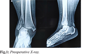
A 60 year old female was brought to emergency department after she sustained an injury to her right ankle after a road traffic accident. On clinical examination she had swelling and deformity of her right ankle. She had tenderness along the distal fibula and over deltoid ligament in the medial aspect. She was diagnosed with type C Weber bimalleolar fracture right ankle [Fig.1]. It was a supination external rotation injury (Lauge Hansen classification). After initial resuscitation and provisional limb stabilisation, she was planned for elective surgery.
After pre-operative planning, the ankle fracture was stabilised as per the principles of AO. The fibula was brought to length and stabilised with distal fibular locking plate. After stabilisation the stress test confirmed the syndesmotic injury. Hence the syndesmotic tightrope was passed through the plate and stabilised. Tightrope comprised of non-absorbable fiber wire suture looped twice through a central holes of anchored cortical metal buttons.
A drill hole was made through all four cortices in a parallel fashion along the transmalleo-lar axis, 2 cm above the ankle joint. A needle containing the pull through suture was then advanced through the drill hole from lateral to medial aspect. Once the suture button crossed the farthest cortex, the button was flipped and attached to the medial tibial cortex, following which the tightrope is compressed by giving traction to the sutures and tied by hand. Intra-operatively syndesmotic disruption is corrected with foot plantigrade and internal rotation while the syndesmotic tightrope is compressed [Fig.2-5].
Post-operatively the patient was allowed full weight bearing mobilisation initially with walker support and followed by full weight bearing with no support. She was reviewed for suture removal on 14th post-operative day and reviewed after three months and one year. She returned to her daily activity after two weeks and was ambulant independently. She had no implant related symptoms.
Discussion
The syndesmosis is injured in mechanisms of severe abduction or external rotation at the ankle joint [8,9] or in cases of proximal fibula fracture with interosseus ligament disruption (Maisonneuve fracture). Surgical management of syndesmotic injury is imperative to prevent ankle instability and secondary osteoarthritis. Surgical fixation of syndesmosis is indicated if there is a severe injury rendering ankle joint unstable under physiological forces [8]. Historically surgical fixation using diastasis screw fixation was the method of choice. However opinions vary widely with regard to type, size, number, position of screws and timing of removal in literature.
Moreover screw fixation has few disadvantages like excessively rigid fixation at normally a mobile joint, extended post-operative non-weight bearing, non-anatomical reduction, revision surgery, screw loosening (20%) and breakage (28%) [7]. Tightrope stabilisation, a non-absorbable fiberwire suture looped twice through center holes of anchored cortical metal button of an ankle syndesmotic injury has been presented as an alternative that has good or improved outcomes and is more physiological than standard screw techniques. The suture resists diastasis of the joint, but under tension allows more movement than a metal screw. Because of the non-absorbable material, a second surgery is not required to remove the tightrope; it is a permanent stabilization system. Prospective studies have demonstrated better American Orthopaedic Foot and Ankle Society scores, earlier return to work, and lower complication rates compared to traditional screw fixation.
Miller et al. [4] demonstrated, in a cadaver model, that a construct with only 2 strands of no. 5 fiberwire suture placed through bony tunnels was equivalent to a single 3.5 mm tricortical screw in resisting a distraction force at the mortise. Jelinak and Porter [9] suggest, following screw fixation, a professional athlete should expect to take between 120 and 180 days to return to sport. Hunt et al. [10] suggested 70-84 days is the likely RTP time frame with their preferred method of suture button fixation. Alex Latham [11] in his case series suggested return to sport is possible within 61 days following syndesmosis injury, provided surgery with tightrope and rehabilitation is uncomplicated.
Forsythe et al. [12] in their cadaveric study supported screw fixation over tightrope as the immediate external rotation force applied after fixation was stable in screw fixation group. But on ideal clinical conditions, the external rotation force is avoided for six weeks to allow syndesmosis healing. In addition currently there are studies evaluating the significance of using two or more tightropes to increase the rigidity of the construct but allowing talofibular motion. There is also a concern that the buttons may pull through the cortex, making the fixation useless. This is especially of concern when the medial button is placed against the metaphyseal cortex. Hence it is important to have at least one of the tightropes through the thicker, more proximal cortical bone.
Despite the above concerns, tightrope offers significant potential advantages over conventional screw fixation. Insertion of the device is simple, both in isolation and in combination with fixation of fibula fractures. The potential for a second operation for implant removal is avoided. In addition, due to the flexibility of the device, the fibula is pulled into the incisura of the tibia as it is tightened, potentially leading to an improved reduction of the syndesmosis
There is currently no evidence as to the optimum time between injury and surgical repair, however, many studies agree that early management provides the best outcome [13,14].
Conclusion
Tightrope fixation offers the potential of syndesmosis stabilization without eliminating normal tibiofibular motion. This may, in turn, lead to better objective ankle motion as well as a decreased subjective stiffness and discomfort making it as a perfect alternative for syndesmotic screw fixation.
Contributors: SK: manuscript writing, patient management; ABG, VK: manuscript editing, patient management; VAN, AM: critical inputs into the manuscript. SK will act as guarantor. All authors approved the final version of this manuscript.
Funding: None; Competing interests: None stated.
References
- Hermans JJ, Beumer A, de Jong TA, Kleinrensink GJ. Anatomy of the distal tibiofibular syndesmosis in adults: a pictorial essay with a multimodality approach. J Anat. 2010;217:633-645.
- Warner SJ, Fabricant PD, Garner MR. The measurement and clinical importance of syndesmotic reduction after operative fixation of rotational ankle fractures. J Bone Joint Surg Am. 2015;97:1935-1944.
- Al-azzani WAK, Sabah T, Paringe V. Evaluation of ankle tightrope syndesmosis fixation. J Foot Ankle Surg Asia Pacific. 2014;1:1-4.
- Miller TL, Skalak T. Evaluation and treatment recommendations for acute injuries to the ankle syndesmosis without associated fracture. Sports Med. 2014;44:179-188.
- Klitzman R, Zhao H, Zhang LQ, Strohmeyer G, Vora A. Suture-button versus screw fixation of the syndesmosis: a biomechanical analysis. Foot Ankle Int. 2010;31:69-75.
- Cottom JM, Hyer CF, Philbin TM. Transosseous fixation of the distal tibiofibular syndesmosis: comparison of an interosseous suture and endobutton to traditional screw fixation in 50 cases. J Foot Ankle Surg. 2009;48:620-630.
- Mulligan EP. Evaluation and management of ankle syndesmosis injuries. Physical Therapy in Sports. 2011;12:57-69.
- Jensen SL, Andresen BK, Mencke S, Nielsen PT. Epidemiology of ankle fractures: A prospective population-based study of 212 cases in Aalborg, Denmark. Acta Orthop Scand. 1998;69:48-50.
- Jelinek JA, Porter DA. Management of unstable ankle fractures and syndesmosis injuries in athletes. Foot Ankle Clin. 2009;14:277-298.
- Hunt KJ, Phisitkul P, Pirolo J. High ankle sprains and syndesmotic injuries in athletes. J Am Acad Orthop Surg. 2015;23:661-673.
- Latham AJ, Goodwin PC, Stirling B, Budgen A. Ankle syndesmosis repair and rehabilitation in professional rugby league players: a case series report, BMJ Open Sport and Exercise Medicine. 2017;3:e000175.
- Forsythe K, Freedman K, Stover M. Comparison of a novel fiberwire-button construct versus metallic screw fixation is a syndesmotic injury model. Foot Ankle Int. 2008;29:49Y54.
- Mak MF, Gartner L, Pearce CJ. Management of syndesmosis injuries in the elite athlete. Foot Ankle Clin. 2013;18:195-214.
- Hsu AR, Garras DN, Lee S. Syndesmotic injuries in athletes. Oper Tech Sports Med. 2014;22:270-281.