Pareshkumar A. Thakkar, Sheila Aiyer, Binoy Shah
From the Department of Pediatrics,
Medical College and S S G Hospital, Vadodara, Gujarat, India.
Corresponding Author:
Dr. Pareshkumar A. Thakkar
Email: pareshthakkar@yahoo.com
Abstract
Jeune syndrome (asphyxiating thoracic dystrophy, ATD) is a rare autosomal recessive skeletal dysplasia characterized by a small, narrow thorax and short limb dwarfism with a considerable neonatal mortality. We report a case of Jeune syndrome admitted at our neonatal ICU. The child had narrow thorax with short limbs. Skeletal survey showed typical findings of horizontal short ribs with irregular and enlarged costochondral junction, small and horizontal clavicles.
|
6go6ckt5b8|3000F7576AC3|Tab_Articles|Fulltext|0xf1ffd84c010000004c00000001000100
6go6ckt5b5idvals|129
6go6ckt5b5idcol1|ID
6go6ckt5b5|2000F757Tab_Articles|Fulltext
Jeune syndrome or asphyxiating thoracic dystrophy (ATD; OMIM 208500) is a rare multisystem autosomal recessive skeletal dysplasia characterized by a narrow thorax, short-limbed dwarfism, renal, hepatic, pancreatic and retinal anomalies. It is estimated to occur in 1 per 1,00,000-1,30,000 live births [1]. We describe a one such case with diagnosis based on clinical and radiographic findings.
Case Report
A full term male child of non-consanguineous parents with a birth weight of 2500 grams was born to a primigravida mother. The child was delivered normally, sustained birth asphyxia and developed respiratory distress soon after birth. There was no history suggestive of antenatal risk factors in mother and family history of similar nature.
Examination revealed full term appropriate for gestational age infant having small bell shaped chest with narrow thorax and short limbs [Fig.1]. The signs of respiratory distress with chest indrawings were present. The cardiovascular system and ophthalmological examination were unremarkable with no facial dysmorphism. Liver function tests were normal except for mild elevation of alkaline phosphatase. Urine examination was negative for proteinuria and hematuria.
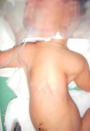
Fig.1: Asphyxiating Thoracic Dystrophy (Jeune Syndrome).
The skeletal survey revealed horizontal clavicles at the level of sixth cervical vertebra, narrow thoracic cavity with widening in lower region, increased density of bone predominantly thorax, vertebrae, horizontal ribs with enlarged costochondral junction, shortened iliac bone with trident appearance of acetabula [Fig.2].
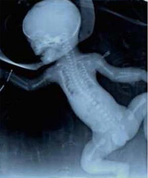
Fig.2: Infantogram of newborn with Asphyxiating thoracic dystrophy.
During his NICU stay, the child was started on oxygen, intravenous fluids and antibiotics for respiratory distress. Subsequently, respiratory distress worsened and child was put on mechanical ventilation. The child expired within 48 hours due to multiorgan failure.
Discussion
Asphyxiating thoracic dystrophy was first described in 1955 by Jeune et al in two siblings with severely narrow thorax [2]. Jeune syndrome is known to be genetically heterogeneous. The inheritance of ATD is autosomal recessive. A locus has been identified on chromosome 15q13 [3], while recently, mutations were found in the IFT80 gene, encoding an intraflagellar protein, in a small subset of ATD patients, who did not have extra skeletal manifestations [4].
Clinically, ATD is characterized by a small, narrow chest and variable limb shortness. Associated congenital abnormalities can be postaxial polydactyly of both hands and/or feet (20%). Typical radiographic findings include a narrow, bell-shaped thorax with short, horizontally oriented ribs and irregular costochondral junctions, elevated clavicles, short iliac bones with a typical trident appearance of the acetabula, relatively short and wide long bones of the extremities, and hypoplastic phalanges of both hands and feet with cone-shaped epiphyses [5,6,7]. The reported case had narrow thorax, elevated clavicles, horizontal ribs, enlarged costochondral junctions, short iliac bones with trident acetabula. The small, narrow thorax often results in respiratory distress and recurrent respiratory infections in the neonatal period and infancy. Ultrasonography findings suggestive of short proximal bones (under 3 percentiles), and a diminished thoracic circumference (although greater than 10 percentiles for the gestational age), at around 23-24 weeks can help in prenatal diagnosis of Jeune’s syndrome in high risk mothers.
The radiographic findings are so typical that distinction from other skeletal dysplasias, except Ellis–van Creveld syndrome, is not difficult. In Ellis–van Creveld syndrome the appearance of the pelvis is indistinguishable, but the involvement of the thorax is less pronounced [6,7].
Jeune syndrome is sometimes compatible with life, although respiratory failure and infections are often fatal during infancy. The severity of thoracic constriction widely varies. For those patients who survive infancy, the thorax tends to revert to normal with improving respiratory function. This suggests that the lungs have a normal growth potential and the respiratory problems are secondary to restricted rib cage deformity [8].
Swiss made Cheap fake rolex uk online store - All perfect replica Rolex watches are all available.
UK 1:1 cheap rolex replica watches uk at discount price including Submariner, Daytona, Datejust, Yacht-Master and so on can be found from this website.
It is important to establish a correct diagnosis both in severe and mild forms since ATD might recur within the same family [9].
References
- Oberklaid F, Danks DM, Mayne V, Campbell P. Asphyxiating thoracic dysplasia: clinical, radiological, and pathological information on 10 patients. Arch Dis Child 1977; 52:758–765.
- Jeune M, Beraud C, Carron R. Dystrophie Thoracique asphyxiante de caractère familial. Arch Fr Pediatr 1955;12:886.
- Morgan NV, Bacchelli C, Gissen P, , Morton J, Ferrero GB, SilengoM et al a locus for asphyxiating thoracic dystrophy, ATD maps to chromosome 15q13. J Med Genet 2003;40:431–435.
- Beales PL, Bland E, Tobin JL et al. IFT80, which encodes a conserved intraflagellar transport protein, is mutated in Jeune asphyxiating thoracic dystrophy. Nat Genet 2007;39:727–729.
- Hennekam RCM, Beemer FM, Gerards LJ, Cats B. Thoracic pelvic phalange dystrophy (Jeune syndrome). Tijdschr Kindergeneeskd 1983;51:95–100.
- Herdman RC, Langer LO. The thoracic asphyxiant dystrophy and renal disease.Am J Dis Child 1968;116:192–201.
- Pirnar T, Neuhauser EBD. Asphyxiating thoracic dystrophy of the newborn. Am J Roentgen 1966;98:358–364.
- de Vries J, Yntema JL, van Die CE, Crama N, Cornelissen EAM, Hamel BCJ. Jeune syndrome: description of 13 cases and a proposal for follow-up protocol. Eur J Pediatr 2010;169:77–88.
- Tüysüz B, Baris S, Aksoy F, Madazli R, Üngür S, Sever L. Clinical variability of asphyxiating thoracic dystrophy (Jeune) syndrome: Evaluation and classification of 13 patients. Am JMed Genet 2009; 149A: 1727-1733.
|