6go6ckt5b8|3000F7576AC3|Tab_Articles|Fulltext|0xf1ff688c310000007b06000001000e00
6go6ckt5b5idvals|2043
6go6ckt5b5|2000F757Tab_Articles|Fulltext
Introduction
Triple X syndrome is also called trisomy X or 47,XXX; sex chromosome anomaly characterized by the presence of an extra X chromosome in a female. Triple X syndrome is first described by Jacob et al. [
1]. An extra copy of X chromosome is generally associated with tall stature, relatively underweight for their height, learning problems, menstrual abnormalities and sometimes with adult premature ovarian failure [
2].
Case Report
A 24-year-old woman, born to non-consanguineous parents, was referred to our institute for the evaluation of primary amenorrhea. She was the third born child in her family, with normal developmental milestones and having body weight of 48 kg and height 146 cm. No stigma of Turner syndrome was noticed. She had bilateral short 4th and 5th toes. Axillary and pubic hairs were absent indicating possible androgen insensitivity. Breast development was Tanner stage-II. Her intelligence quotient was within the normal range. A preliminary diagnosis of primary amenorrhea was made.
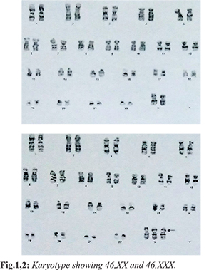
Ultrasound of pelvic region showed small uterus of 51×21×13 mm size. Endometrium was 1.2 mm thick. Her both ovaries were rudimentary measuring 12×5 mm and 13×6 mm respectively. However, the vagina was well visualized. Her luteinizing hormone (LH) was (51.84 mIU/mL) and follicle stimulating hormone (FSH) was (119.58 mIU/mL) suggesting premature ovarian insufficiency. Cytogenetic analysis was carried out by GTG banding on cultured peripheral blood lymphocytes by standard technique. In the present study, 50 metaphase plates were analysed, which revealed mosaicism of 46,XX/47,XXX chromosomal constitution with 90% of the cells showing 46,XX
[Fig.1] and 10% of the cells exhibiting 47,XXX
[Fig.2]. The index patient was having short stature. The woman was 146 cm tall considering her father and mother’s height were 170 cm and 157 cm respectively with mid parenteral height was 165.5 cm. According to the Indian Academy of Paediatrics (IAP) growth charts her height is below 3rd centile [
3].
In summary based upon the clinical features, hormonal assay and cytogenetics, a formal diagnosis of primary ovarian failure (POF) with mosaic triple X syndrome was made. The first association of triple X syndrome with POF is described by Jacobs et al. in 1959 [
1]. Although non-mosaic 47,XXX karyotypes are the most frequent; mosaicism occurs in approximately 10% of cases with many combinations such as 46,XX, 47,XXX, or 48,XXXX [
4].
Discussion
POF may be the presenting symptom in up to 10% of cases of primary amenorrhea [
5]. Primary ovarian failure as a presentation of Triple X syndrome constitutes only 3.8% of cases in literature [
6]. Genetic cause constitutes 33% of the POF which involves mostly the X chromosome [
7]. Considering the poor breast development (Tanner II) and amenorrhea, she was started initially with ethinyl-oestradiol 10 µg daily. It was planned to start with low dose estrogens and progesterone oral contraceptive pills; till she starts regular menstruation or she attained proper Tanner breast staging. Future plan for management of this case was to get her counselled by a qualified psychologist about the permanent infertility that she will have and plan for adoption if she wishes to marry in future.
Table 1 depicts 47,XXX-triple X syndrome patients with premature ovarian failure reported in the literature.
Most of the subjects presented in literature were having normal stature along with no mental abnormality. Our case was different from the other being short stature with mosaicism of 46,XX/47,XXX with normal scholastic performance. She presented with primary amenorrhea with poor secondary sexual character and was managed appropriately with low dose oral contraceptive pills.
Conclusion
Our case is unique being presented as short stature. It also stresses upon the utility of chromosomal karyotype in women presenting with primary amenorrhea with premature ovarian failure for diagnosis and appropriate management. A multidisciplinary approach is needed for proper treatment.
Contributors: KKB: Patient management and preparation of draft manuscript; JB: patient management and critical inputs into the manuscript; DH: manuscript revision. KKB acted as study guarantor. All authors approved the final version of the manuscript and are responsible for all aspects of the study.
Funding: None; Competing interests: None stated.
References
- Jacobs P, Baikie AG, Court Brown WM, Macgregor TN, Maclean N, Harnden DG. Evidence for the existence of the human “super female.” The Lancet. 1959;26:423-425.
- Otter M, Schrander-Stumpel CT, Curfs LM. Triple X syndrome: a review of the literature. Eur J Hum Genet. 2010;18:265-271.
- Khadilkar V, Yadav S, Agrawal KK, Tamboli S, Banerjee M, Cherian A, et al. Revised IAP growth charts for height, weight and body mass index for 5- to 18-year-old Indian children. Indian Academy of Pediatrics Growth Charts Committee, Indian Pediatr. 2015;52:47-55.
- Tartaglia NR, Howell S, Sutherland A, Wilson R, Wilson L. A review of trisomy X (47, XXX). Orphanet J Rare Dis. 2010;5:8.
- Ebrahimi M, Akbari AF. Pathogenesis and causes of premature ovarian failure: An update. Int J Fertil Steril. 2011:54-65.
- Goswami R, Goswami D, Kabra M, Gupta N, Dubey S, Dadhwal V. Prevalence of the triple X syndrome in phenotypically normal women with premature ovarian failure and its association with autoimmune thyroid disorders. Fertil Steril. 2003;80:1052-1054.
- Torrealday S, Kodaman P, Pal L. Premature ovarian insufficiency - an update on a recent advance in understanding and management. F1000Res. 2017;29:2069.
- Chandana C, Venkatesh S. Triple X syndrome woman presenting as premature ovarian failure. Int J Infertil Fetal Med. 2013;4:96-98.
- Kodandapani S, Pai MV, Nambiar J, Moka R. Premature ovarian aging in primary infertility: Triple X syndrome. J Hum Reprod Sci. 2011;4:153-154.
- Villanueva AL, Rebar RW. Triple-X syndrome and premature ovarian failure. Obstet Gynecol. 1983;62:70S-73S.