6go6ckt5b8|3000F7576AC3|Tab_Articles|Fulltext|0xf1ff186003000000f601000001000600
6go6ckt5b5idvals|214
6go6ckt5b5idcol1|ID
6go6ckt5b5|2000F757Tab_Articles|Fulltext
Introduction
Esophageal atresia (EA) with Tracheoesophageal fistula (TEF) repair is an emergency which needs intubation of trachea with tube tip beyond fistula and controlled ventilation as trachea is communicating with oesophagus [
1]. Unexpected subglottic stenosis in patients of TEF repair is challenging case for the anaesthesiologist as it is difficult to diagnose this condition with preoperative predictors of difficult airway, more so in a neonate. General anaesthesia with endotracheal intubation can lead to damage to laryngeal cartilage and tracheal perforation [
2]. In such cases, repeated and forceful attempts at intubation may lead to oedema of already compromised airway [
3].
Case Report
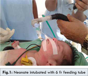
A 1 day old full term male baby, weighing 2.2 kg presented as a case of esophageal atresia with tracheoesophageal fistula. Pre-operative assessment of the patient showed stable vitals. These included heart rate-140/min, respiratory rate-40/min, SpO2-98% on room air. Under monitoring patient was pre-medicated with injection atropine 0.01 mg/kg and induced with sodium thiopentone and succinylcholine 1.5 mg/kg. Subglottic stenosis was suspected in view of endotracheal intubation not being achieved even with smallest sized 2 mm ID endotracheal tube. Again after pre-oxygenation patient was intubated with 6 fr feeding tube cut short at 38.5 cm so as to match with length of 2 mm ID endotracheal tube and connector of 3.5 mm ID ETT attached, to aid proper connection with Jackson rees’ modification of Ayres’ T-piece [Fig.1].
Patient was maintained on 50% oxygen and 50% air and halothane, injection atracurium was given for muscle relaxation. Right postereolateral thoracotomy and primary esophageal anastomosis was done along with bronchoscopic dilatation of the subglottic stenosis. At the end of procedure, patient was reversed but was not extubated and kept on T-piece with continuous monitoring [Fig.2,3]. Post operatively patient became fully conscious, had good respiratory effort was moving all limbs and maintaining saturation of 98%. After suctioning oral cavity, patient was extubated.
Patient was shifted to neonatal intensive care unit for 7 days, feeds started and subsequently discharged.
Discussion
Subglottic stenosis (SGS) is narrowing of the subglottic airway. It extends from lower surface of the true vocal cords to the lower surface of cricoids cartilage. The subglottic airway is considered the narrowest part of airway as it is complete, non expandable and non pliable tube and does not have posterior membranous or muscular section like trachea and larynx [
4]. At birth infant larynx is about 1/3rd of size of adult larynx however, it is proportionately larger than that of adult compared to remaining tracheobronchial tree. The term SGS implies a narrowing that can be congenital or acquired. The current incidence of neonatal subglottic stenosis is less than 2.0% [
5]. In congenital SGS, generally conservative approach is employed while severe form requires intervention in the form of endoscopic or external reconstruction [
6]. Myer and Cotton gave staging system and described 4 grades of obstruction: Grade 1: 0-50% of the lumen obstruction; Grade 2: involvement of 51-70% of the lumen; Grade 3: obstruction of 71- 99% of lumen; Grade 4: 100% obstruction of lumen [
7]. Supraglottic devices are generally indicated in management of SGS but in our case, it was not possible to manage the patients as the goal of intubation was to have the tip of endotracheal tube beyond the TEF fistula but proximal to carina [
8,
9]. It is necessary to intubate patient, as with supraglottic devices air will leak through fistula, desaturation may result due to atelectasis caused by lung retraction. Tracheostomy was not feasible in our case. T Asai reported a similar case report in a infant in which size 2 mm ID ETT tube failed to pass, subglottic stenosis was dilated using 7 fr percutaneous transluminal coronary angioplasty catheter with balloon under fluoroscopy and passing size 3 mm ETT through the glottis and surgery was performed after 2 days. But same was not possible in our case due to lack of fluoroscopy, although bronchoscopic dilatation was done in our case. The need of effective airway resulted in use of feeding tube which was having lesser diameter than smallest size 0.2 mm endotracheal tube.
Conclusion
The patients of oesophageal atresia with respiratory distress should be evaluated preoperatively to rule out unexpected complications like subglottic stenosis. This surgical emergency can be managed in unique manner by a newer technique of using 6 fr feeding tube which has outer diameter lesser than that of 2 mm ID endotracheal tube for successfully carrying out surgical procedure.
Acknowledgements
We would like to acknowledge Dr. Ritika Basnotra in helping with the design, concept and preparation of this manuscript.
References
- Gayle JA, Gomez SL, Baluch A, Fox C, Lock S, Kaye A. Anaesthetic consideration for the neonate with tracheoesophageal fistula. MEJ Anesth. 2008;19:1241-1254.
- Norris BK, Schweinfurth JM. Aretenoid dislocation: An analysis of contemporary literature. Laryngoscope. 2011;121:142-146.
- Mort TC. Emergency Tracheal Intubation: Complications Associated with Repeated Laryngoscopic Attempts. Anesth Analg. 2004;99:607-613.
- Eckel HE, Koebke J, Sittel C, Sprinzl GM, Pototschnig C, Stennert E. Morphology of the human larynx during the first five years of life studied on whole organ serial sections. Ann Otol Rhinol Laryngol. 1999;108:232–238.
- Walner DL, Loewen MS, Kimura RE. Neonatal subglottic stenosis—incidence and trends. Laryngoscope. 2001;111:48-51.
- Cotton RT. Pediatric laryngotracheal stenosis. J Pediatr Surg.1984;19:699-703.
- Myer CM, O’Connor DM , Cotton RT. Proposing grading system for subglottic stenosis based on endotracheal tube size. Ann Otol Rhinol Laryngol. 1994;103:319-323.
- Andropoulos DB, Rowe RW, Betts JM. Anaesthetic and surgical airway management during Tracheoesophageal fistula repair. Pediatr Anaesth. 1998;8:313-319.
- Yavascaoalu B, Tokat O, Basagan EM, Kaya FN, Erisen L, Kutlay O. The use of the laryngeal mask airway in children with subglottic stenosis. J Int Med Res. 2001;29:541-545.