6go6ckt5b8|3000F7576AC3|Tab_Articles|Fulltext|0xf1fff882050000009502000001000c00
6go6ckt5b5idvals|299
6go6ckt5b5|2000F757Tab_Articles|Fulltext
Introduction
In females, the round ligament is attached to the uterus near the origin of the fallopian tube and a small evagination of parietal peritoneum accompanies the round ligament through the inguinal ring into the inguinal canal [
1]. This small evagination of parietal peritoneum is the canal of Nuck in the female, homologous to the processus vaginalis in the male. It was described by Dutch anatomist Anton Nuck in 1691[
2]. The canal of Nuck normally undergoes complete obliteration during the first year of life. Failure of complete obliteration results in either an indirect inguinal hernia or a hydrocele (cyst) of the canal of Nuck [
1,
3].
Endometriosis is described as the implantation of endometrial tissue outside the uterus affecting 1-2% of women [
4]. It usually involves ovaries and peritoneum [
5] although rare cases have been reported in lungs, rectum, vagina, subcutaneous tissue and inguinal canal [
6]. The patent canal of Nuck, by virtue of creating a communication between the peritoneal cavity and inguinal canal, helps in retrograde movement of endometrial tissue into the extraperitoneal space [
2]. Such endometrial deposits in cyst of canal of Nuck can present as an inguinal swelling. A cyst of the canal of Nuck is a rare condition and occurrence of endometriosis in it is yet rarer.
Case Report
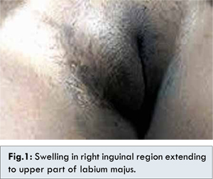
A 29 year old woman presented with a swelling in her right groin which had been gradually growing in size since 6 months with no other complaint. Her medical, obstetrical and menstrual histories were unremarkable. There was no alteration in the size of lump during menses or on lying supine. Examination revealed a firm, mobile, non-tender, irreducible mass of size 4x3 cm having smooth surface in right inguinal region extending to upper part of labium majus with no alteration in size on Valsalva manoeuvre [Fig.1]. Ultrasonography revealed a well-defined hypoechoic lesion with internal echoes.
On exploration of inguinal region, a firm sub-aponeurotic lump continuous with round ligament was found. On dissecting the lump, its interior revealed several small loculi containing scanty yellowish-brown fluid. Proximally, the lumen was occluding and merging with round ligament. The lump was excised en-block and subjected for histopathological examination. Histopathological examination demonstrated a loculated cyst of canal of nuck with deposition of endometrial tissue.
Discussion
The canal of Nuck is a small protrusion of peritoneum in inguinal canal of females that corresponds to the processus vaginalis in the males. The processus vaginalis, in both the sexes, normally undergoes obliteration during the first year of life [
1]. If obliteration fails in the distal portion of the canal, a sac containing serous fluid remains, the so called hydrocele or cyst of canal of Nuck [
1,
3]. A cyst of canal of Nuck can result from either a persistent patent processus vaginalis with peritoneal communication or with proximal obliteration at the deep ring with over-secretion and under-absorption in distal segment. In such cases, the canal provides the most likely pathway for endometrial tissue to implant into inguinal canal [
2]. It has been postulated that extra-pelvic endometriosis that is distant to the uterus tends to lose its hormonal receptors and response, hence the lack of cyclical symptoms [
2]. Clinically, the cyst of the canal of Nuck manifests as a painless swelling in the inguinal area and labium majus.
The most common differential of cyst (hydrocele) of the canal of Nuck is the inguinal hernia, and both can coexist in about one-third of patients. Other differentials include incarcerated hernias, abscesses, vascular abnormalities, and cystic-appearing tumors. Ultrasound is the imaging modality of choice in the assessment of groin lesions as it usually resolves most of these differentials with careful technique and application of colour Doppler [
7]. Exploration of inguinal canal and total excision of cyst is the definitive treatment being both therapeutic as well as diagnostic. This case illustrates the importance of considering the cyst of canal of Nuck, though rare, as a potential differential diagnosis in young females presenting with inguinal swellings.
A thorough clinical examination of the patient revealed a firm, non-tender, non-reducible and mobile mass, with a smooth surface, from the inguinal canal to the labium majus. It raised a suspicion of a cyst in the canal of Nuck. Ultrasound imaging was used as a modality to confirm the diagnosis. Surgical exploration and cyst excision was the definitive treatment. Histopathological examination of the cyst revealed deposits of endometrial tissue.
Conclusion
A rare presentation of endometriosis in cyst of canal of Nuck is reported here. A cyst (hydrocele) of the canal of Nuck though rare should be considered in the differential diagnosis in females presenting with an inguinal swelling. Establishing a definitive diagnosis on clinical examination is challenging, radiological imaging and guided aspiration cytology may assist in diagnosis but surgical exploration and histopathological examination is critical for final diagnosis and treatment.
References
- Anderson CC, Broadie TA, Mackey JE, Kopecky KK. Hydrocele of the canal of Nuck: ultrasound appearance. Am Surg. 1995;61:959-961.
- Shadbolt CL, Heinze SBF, Dietrich RB. Imaging of the groin masses: inguinal anatomy and pathological conditions revisited. Radiographics. 2001;21:S261-S271.
- Schwartz A, Peyser MR. Nuck’s hydrocele. Int Surg. 1975;60:91-92.
- Wellbery C. Diagnosis and Treatment of Endometriosis. Am Fam Phys. 1999;60:1753-1762.
- Seli E, Berkkanoglu M, Arici A. Pathogenesis of endometriosis. Obstet Gynec Clin North Am. 2003;30:41-61.
- Strasser EJ, Davis RM. Extraperitoneal inguinal endometriosis. Am Surg.1977;43:421-422.
- Edmund Soh, Kenneth Sheah, Keh Oon Ong. Hydrocele of the canal of Nuck: ultrasound and MRI findings. Reports in Medical Imaging. 2011:4:15-17.