Introduction
Mucopolysaccharidosis syndrome represents a family of rare inherited lysosomal storage disorders which lead to deficiency in enzymes necessary for glycosaminoglycan degradation. This results in increase in the concentration of tissue glycosaminoglycan. Cardiovascular involvement is reported in approximately 50-90% of cases and is characterized by valvular thickening, myocardial hypertrophy, and fibrosis of the chordae tendineae [
1]. Among these, mitral valve pathology is most frequently observed, often coexisting with aortic valve abnormalities and left ventricular hypertrophy. We describe a case of mucopolysaccharidosis presenting with severe mitral stenosis, culminating in the necessity for mitral valve replacement.
Case Report
A 12-year-old female presented with complaints of exertional dyspnea persisting for two months, accompanied by pedal edema. The patient had normal intellectual development and an unremarkable past medical history. On examination, she exhibited characteristic morphological features including short stature, a short neck, and skeletal irregularities [Fig.1].
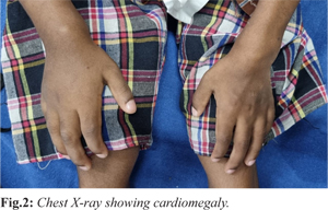
Transthoracic two-dimensional echocardiography (2D ECHO) revealed severe mitral stenosis with mild mitral regurgitation, a single papillary muscle, transmitral gradients of 34/19 mmHg, and evidence of right ventricular dysfunction. Radiographic evaluation demonstrated cardiomegaly [Fig.2]. Ultrasonography of the abdomen and pelvis showed bilaterally small to borderline kidneys. A mucopolysaccharidosis (MPS) spot test yielded a positive result, and MPS electrophoresis detected a trace chondroitin sulfate band, confirming the diagnosis.
The patient was scheduled for corrective surgery. Intraoperative findings included cardiomegaly with severe calcific mitral stenosis, a fixed valvular orifice, fusion of the subvalvular apparatus, and moderate tricuspid regurgitation [Fig.3]. The surgical procedure entailed mitral valve replacement using a 23 mm St. Jude Medical mechanical prosthesis and tricuspid valve repair reinforced with Teflon felt. The immediate postoperative course was uneventful. A repeat 2D ECHO on the third postoperative day demonstrated improved right ventricular function and reduced mitral valve gradients of 11/4 mmHg.
Histopathological examination of the excised mitral valve tissue revealed mucopolysaccharide infiltration consistent with MPS-related valvular pathology. The patient was discharged in a hemodynamically stable condition. During the two-year postoperative follow-up period, she remained asymptomatic with stable cardiac function.
Discussion
MPS is a family of hereditary disorders of proteoglycan degrading enzymes. Patients with MPS have intracellular accumulation of acid mucopolysaccharides and glycolipids in various tissues and excessive urinary excretion of dermatan, chondroitin and heparin sulphates. Each type has distinct clinical features and absent or insufficient lysosomal enzyme activity. The global incidence of MPS is estimated to be 1-17 cases per 1,00,000 live births and there is a potential influence of geographic and ethnic factors on the distribution of MPS types [
3].
Most prominent cardiac feature in MPS is progressive valvular disease which was first described in the year 1960. The impact of mucopolysaccharidoses (MPS) on valvular interstitial cells, resulting in the activation and development of "Hurler" cells, along with an inflammatory response and macrophage activation, contributes to valve thickening and compromised leaflet mobility in MPS patients. This process is reported to affect a significant percentage of MPS patients, ranging from 60% to 90% [
4].
Mitral regurgitation is more common than mitral stenosis in MPS. There have been only a few case reports of mitral stenosis [
5]. Mitral stenosis caused by mucopolysaccharide infiltration of the valve cusps was first recorded in a case of atypical gargoylism by Vanace, et al. in 1960. Life expectancy in MPS depends on the severity of symptoms [
6]. Without treatment, severely affected individuals survive only till late childhood or adolescence. Milder forms of the disorder usually live into adulthood. Increased mortality in MPS is often due to restrictive lung disease, upper and lower respiratory infections and advanced valvular heart disease [
7].
Mitral valve replacement can be challenging due to small annulus and often leading to patient prosthesis mismatch. Selecting a prosthesis with the largest effective orifice area is crucial for minimizing postoperative pressure gradient. Only a few cases of successful mitral valve repair have been reported till date.
Conclusion
Surgical valve replacement becomes necessary for MPS patients facing progressive, severe valvular disease despite systemic treatment advances. Precise multidisciplinary pre-operative planning, personalized solutions, and an experienced clinical team are crucial to handle such cases. Reporting such cases is vital to improve recognition, guide management, and build evidence for future therapeutic decisions.
Contributors: CLA: concept and manuscript writing; PSSG: manuscript editing and literature search. CLA will act as a study guarantor. All authors approved the final version of this manuscript and are responsible for all aspects of this study.
Funding: None; Competing interests: None stated.
References
- Braunlin E, Wang R. Cardiac issues in adults with the mucopolysaccharidoses: current knowledge and emerging needs. Heart. 2016;102(16):1257-1262.
- V Fesslova, P Corti, G Sersale, A Rovelli, P Russo, S Mannarino, et al. The natural course and the impact of therapies of cardiac involvement in the mucopolysaccharidoses. Cardiol Young. 2009;19(2):170-178.
- Khan SA, Peracha H, Ballhausen D, Wiesbauer A, Rohrbach M, Gautschi M, et al. Epidemiology of mucopolysaccharidoses. Mol Genet Metab. 2017;121(3):227-240.
- Braunlin E, Tolar J, MacKey-Bojack S, Masinde T, Krivit W, Schoen FJ. Clear cells in the atrioventricular valves of infants with severe human mucopolysaccharidosis (Hurler syndrome) are activated valvular interstitial cells. Cardiovasc Pathol. 2011;20(5):315-321.
- Semenza GL, Pyeritz RE. Respiratory complications of mucopolysaccharide storage disorders. Medicine (Baltimore). 1988;67(4):209-219.
- Vanace, PW, Friedman S, Wagner BM. Mitral stenosis in an atypical case of gargoylism: a case report with pathologic and histochemical studies of the cardiac tissues. Circulation. 1960;21:80-89.
- Ireland MA, Rowlands DB. Mucopolysaccharidosis type 4 as a cause of mitral stenosis in an adult. Br Heart J. 1981;46(1):113-115.