6go6ckt5b8|3000F7576AC3|Tab_Articles|Fulltext|0xf1ff6cd4250000006407000001000900
6go6ckt5b5idvals|901
6go6ckt5b5|2000F757Tab_Articles|Fulltext
Introduction
Chest wall tuberculosis (TB) poses a diagnostic and therapeutic challenge [
1]. It constitutes 1-5% of all cases of musculoskeletal tuberculosis representing 1-2% of TB overall [
2]. Extra-pulmonary tuberculosis represents 15 to 20% of the cases of tuberculosis affections [
3-
5]. Chest wall tuberculosis is diagnosed late due to non-specific signs and symptoms and involvement of sites such as sternal margins and rib shafts. It may present as a painless cystic mass similar to a cold abscess or sometimes as a firm, moving tissue mass.
Case Report
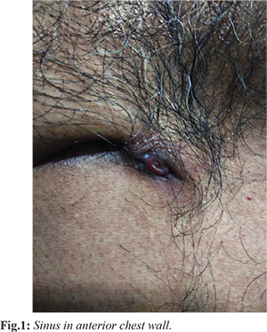
A 45 year old male presented to surgery out-patient department with complain of gradually enlarging soft tissue mass in the right upper anterior thoracic wall. It was first noticed one year back. He was a chronic smoker and had no pulmonary symptoms. There was no history of any trauma or heavy weight lifting. Rest of the medical history of the patient was unremarkable. On examination, the mass was 2×2 cm painless, firm, mobile and well defined. There was no sign of inflammation but the overlying skin showed sinus [Fig.1]. The standard chest X-ray showed no abnormalities. The chest computer tomography performed with low thoracic slices showed homogenous cystic area. No parenchymal lesions were noted. Fine needle aspiration showed few degenerated epithelioid cells along with abundant muscle bundle and hemorrhage suggesting chronic granulomatous disease. Further work up was done and biopsy was done for histo-pathological examination. Histopathology report gave the final diagnosis of chest wall tuberculosis. Based on the context of the clinical and radiological aspects, the fact that the mass was localized, the surgical treatment was indicated. The indication of surgical treatment decided upon diagnostic and therapeutic purposes, and the treatment was initiated. The patient underwent surgical resection of a mass and was given anti-tubercular drug therapy. His post-operative course was uneventful and on follow up three months after completion of anti-tubercular therapy he was stable and asymptomatic.
Discussion
Tuberculosis is a serious infectious disease which mainly affects lungs. The bacteria causing tuberculosis spreads from one person to another through tiny droplets released into the air via coughing or sneezing, lungs being the main target. When the bacteria remains in the body in an inactive state and cause no symptoms, it is termed as latent TB or inactive TB or TB infection. This stage is not contagious. It can turn into active TB, so treatment is important for the person with latent TB to help control the spread of TB. Due to non-specific sign and symptoms, the diagnosis may be delayed. In endemic areas possibility of tuberculosis should be ruled out as early as possible in a patient presenting as pyogenic abscess. Pleura and mediastinal or chest wall lymph nodes must be examined. Rib destruction may or may not be present. Tuberculosis is second only to metastatic malignancies as a cause of destructive lesions of the ribs.
Chest wall tuberculosis may occur by hematogenous dissemination associated with activation of a dormant tuberculous focus or direct extension from a lymphadenitis of chest wall. Tuberculous bacilli invade the pleural space and set up a local or widespread pleuritis; some bacilli transport from the pleural space to the parasternal (or posterior intercostal) lymph nodes; these nodes become caseous and rupture; necrotic and caseous material burrows anteriorly (or posteriorly) to form a cold abscess in the chest wall. Delay in diagnosis and treatment may lead to discharging sinus in chest wall tuberculosis. Fine needle aspiration is important to establish the diagnosis and to exclude other infectious diseases or malignancy. Drainage and debridement procedures adjunct to anti-tuberculous therapy is needed for the rapid alleviation of symptoms and clinical recovery in the treatment of chest wall tuberculosis [
6,
7]. WHO recommends a standard 6-month regimen, according to clinical presentation, bacillary load and response to anti-tubercular therapy, the treatment can be extended up to 9-12 months [
8].
Conclusion
Anti-tuberculous medical treatment and adjunctive surgery are quite effective in chest wall tubercular cases.
Contributors: JA: literature search and manuscript preparation; MA, AH: manuscript review and manuscript editing; MA, NK: patient monitoring and manuscript review. AH will act as a study guarantor. All authors approved the final version of this manuscript.
Funding: None; Competing interests: None stated.
References
- Gaude GS, Reyas AK. Tuberculosis of chest wall without pulmonary involvement. Lung India. 2008;25:135-137.
- Morris BS, Maheshwari M, Chalwa A. Chest wall tuberculosis: A review of CT appearances. Br J Radiol. 2004;77:449-457.
- Morris BS, Varma R, Garg A, Awasthi M, Maheshwari M. Multifocal musculoskeletal tuberculosis in children: appearances on computed tomography. Skeletal Radiol. 2002;31:1-8.
- Roy HR. Chest wall tuberculosis. Int J Surg Pakistan. 2004;19(2):82-83.
- Ozkara S, Kilicaslan Z, Ozturk F, Seymenoglu S, Erdogan AR, Tellioglu C, et al. Tuberculosis in Turkey with regional data. Toraks Dergisi. 2002;3:178-187.
- Paik HC, Chung KY, Jeong HK, Dae HM. Surgical treatment of tuberculous cold abscess of the chest wall. Yonsei Med J. 2002;43(3):309-314.
- Kuzucu A, Soysal O, Gunen H. The role of surgery in chest wall tuberculosis. Interactive Cardiovascular and Thoracic Surg. 2004;3(1):99-103.
- WHO Stop TB Department Strategy and framework for effective tuberculosis control. Treatment of tuberculosis: Guidelines for national programs 2003 Geneva World Health Organization. Available at: https://apps.who.int/iris/bitstream/handle/10665/67890/WHO_CDS_TB_2003.313_eng.pdf;jsessionid= BDFAC4FE597CFBE446E6D638C7854895?sequence=1. Accessed on August 10, 2018.