6go6ckt5b8|3000F7576AC3|Tab_Articles|Fulltext|0xf1ff64a42d0000007109000001000100
6go6ckt5b5idvals|997
6go6ckt5b5|2000F757Tab_Articles|Fulltext
Introduction
Peripheral intravenous (IV) cannulation is a common procedure used for obtaining intravas-cular access. It is a routine bedside intervention performed by doctors, nurses and paramedical staff. Peripheral IV cannulas, invaluable in administering IV drugs, fluids and other adjuncts are not without potential complications. Hematomas, thrombophlebitis, cannula related infections, extravasation, injury to arteries, nerves, tendons which are within close proximity to the vein being cannulated and intravascular fracture of the cannula are some of these. Here we report a case of an intravascular fracture of an IV cannula, which is a rare but serious complication of peripheral cannulation.
Case Report
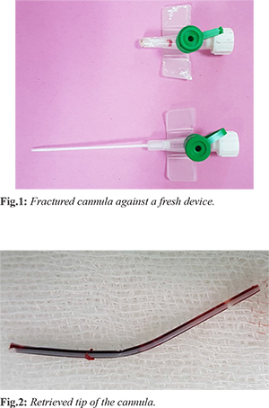
A 61 year old diabetic patient who had kidney transplantation 2 years back was admitted to the surgical unit at National Institute for Nephrology Dialysis and Transplant with a diabetic foot infection. On admission he had stable vitals with a total white cell count of 18×103 /µL (4-10). The C reactive protein level was 221 mg/L (0-6) and serum creatinine was 95.51 µmol/L (60-120). He had a functioning brachio-cephalic arterio venous fistula on his right arm, therefore the left upper limb was cannulated and IV antibiotics were commenced pending wound debridement. At the operating theatre, a 23G IV cannula was inserted to his right external jugular vein (EJV) for IV access as there were no suitable limb veins. Wound debridement was done and IV antibiotics were continued in the ward using the same neck cannula. Three days later when this cannula was being removed it was noted that the tip was missing [Fig.1]. Palpation of the neck revealed some induration along the right EJV which was thought to be caused by the missing piece of cannula left inside the vein. The patient was taken to the theatre and under local anesthesia and the suspicious area of the neck was explored. The EJV was identified, a venotomy made and the cannula tip retrieved [Fig.2]. The vein was ligated. The patient made an uneventful recovery and was discharged on oral antibiotics with follow-up for the foot wound.
Discussion
Peripheral IV cannulation is a common procedure. Approximately 60% of all patients treated by a healthcare system will have an IV cannula inserted for varying purposes [
1]. Among the complications of peripheral venous cannulation, intravascular fracture of the device is an infrequent complication. Its exact incidence is unknown and the literature is limited to case reports.
The broken off piece of cannula within a vein will act as an intravascular foreign body which can embolize. Embolism of broken cannula segments may result in sepsis, endocarditis, cardiac perforations and arrhythmias [
2]. Repeated attempts at insertion, poor quality materials used for device manufacturing and keeping the cannula in situ for prolonged periods have been cited as reasons for breakage of these devices [
3]. Reinsertion of the introducer needle into the cannula during repeated attempts at cannulation can damage the plastic tip of the device. This can lead to complete fracture or partial damage to the tip. A partially damaged cannula can completely transect at the time of its removal [
2]. Leaving an IV cannula in situ for more than 48 hours should be avoided [
4].
Fracture of a cannula may be evident immediately upon its removal or it may present late with local symptoms such as pain and swelling of the affected extremity or symptoms attributable to distal embolization [
3]. Once a cannula fracture is diagnosed the major dilemma is to localize the missing piece. If it is palpable as in our patient exploration without imaging can be attempted. If it is not, blind exploration is likely to be unsuccessful. As almost all of these devices are made of plastic material they are radiolucent, so plain X ray is not helpful. Ultrasound and CT have been used successfully in the literature for locating these intravascular forging bodies [
3,
5]. Once localized by imaging, surgical removal is advisable in order to avoid serious complications.
Proper training and technique can prevent potential complications associated with peripheral cannulation. Reintroduction of the needle into a partially inserted cannula should be avoided. Once inserted, the cannula insertion site should be regularly monitored. At removal the cannula should be checked for its completeness. If a piece of the cannula is missing some recommend an immediate proximal tourniquet to prevent migration of the missing segment until it is localized and removed [
1,
5].
Conclusions
Peripheral IV cannulation is a common procedure invaluable in day to day clinical practice, but healthcare workers should be aware of its associated complications. To avoid these, proper training and correct technique in insertion, adequate surveillance during device maintenance and removal are mandatory.
Contributors: RMTMG: manuscript writing, literature review and patient management; NG: manuscript editing, and patient management; RMTMG will act as a study guarantor. Both authors approved the final version of this manuscript and are responsible for all aspects of the study.
Funding: None; Competing interests: None stated.
References
- Lavery I, Smith E. Peripheral vascular access devices: risk prevention and management.Br J Nurs. 2007;13-2008:916:1378-80, 1382-3.
- Arun O, Bahar OC, Gunduz E, Mehmet Oc, Duman A. Fracture of an intravenous cannula in the vein due to reinsertion of the guide needle: A case report. J Cardiovasc Surg. 2014;2:28-29.
- Singh A, Kaur A, Singh M, Kau S. CT guided removal of iatrogenic foreign body: A broken intravenous cannula. J Clin Diagn Res. 2015;9:PD28-PD29.
- Jayanth BS, Bhandari PM. Fracture of an intravenous cannula in the vein during removal: A Case Report. Nitte University J Health Science. 2016;6:79-81.
- Baliga K, Elamurugan S. Iatrogenic complications: a line’s share. International Surg J. 2016;3:2250-2253.