Introduction
Kabuki syndrome (KS) is a rare congenital disorder, first described independently by Niikawa and Kuroki in 1981, with an estimated prevalence of approximately 1 in 32,000 to 1 in 86,000 live births [
1]. It is characterised by distinctive facial features, developmental delay, skeletal abnormalities, intellectual disability, and variable multisystem involvement, including cardiac, renal, auditory, and immunological abnormalities [
1,
2].
From an anaesthetic perspective, children with KS may pose unique perioperative challenges because of craniofacial and airway abnormalities, including micrognathia, cleft or high-arched palate, hypotonia, cervical spine abnormalities, and congenital cardiac disease [3]. These anatomical variations may complicate airway assessment and management and may occasionally reveal unexpected structural abnormalities during airway instrumentation. We present the perioperative management of a child with KS undergoing planned adenoidectomy, in whom an undiagnosed submucous cleft palate and posterior septal defect were unexpectedly identified intraoperatively. This case highlights the importance of careful airway evaluation and awareness of potentially occult craniofacial abnormalities in children with KS.
Case Report
A 9-year-old female child with a known diagnosis of Kabuki syndrome (KS) presented with bilateral nasal discharge and decreased hearing for several months. She was scheduled for adenoidectomy with bilateral grommet insertion under general anaesthesia. She was born full-term with a birth weight of 2.5 kg and required admission to the neonatal intensive care unit for 5 days following meconium aspiration. Her developmental milestones were delayed, and she subsequently developed bilateral hearing loss and stammering speech, consistent with the neurodevelopmental and otologic manifestations associated with KS.
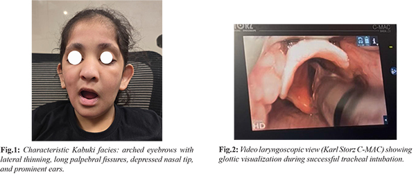
Preanaesthetic evaluation revealed characteristic Kabuki facies, including arched eyebrows with lateral thinning, long palpebral fissures, a depressed nasal tip, and prominent ears [Fig.1]. Airway examination revealed a Mallampati Grade II airway, high-arched palate, adequate mouth opening, and normal neck mobility. No clinically evident cardiovascular or respiratory abnormality was detected. The child was classified as American Society of Anesthesiologists (ASA) Physical Status II. In the operating room, standard ASA monitoring, including electrocardiography, pulse oximetry, and non-invasive blood pressure monitoring, was instituted. Intravenous induction was performed using standard paediatric doses of anaesthetic agents, and mask ventilation was achieved without difficulty. Considering the underlying syndromic diagnosis and the potential for an anatomically difficult airway, neuromuscular blockade was achieved with intravenous rocuronium. Tracheal intubation was subsequently performed successfully using a Karl Storz C-MAC video laryngoscope [Fig.2].
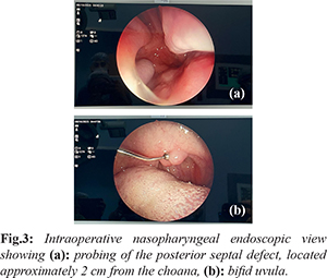
During the intraoperative ENT examination, several previously undiagnosed anatomical abnormalities were identified. These included a bifid uvula and a submucous cleft palate detected on palpation. A posterior septal defect, approximately 2 cm from the choana, was also noted, along with a soft torus tubarius with decreased elasticity [Fig.3]. The adenoids were Grade 1. In view of the occult palatal defect and the associated risk of postoperative velopharyngeal insufficiency following adenoidectomy, the planned surgical procedure was reconsidered. Consequently, adenoidectomy was abandoned, and only bilateral grommet insertion was performed.
The intraoperative course remained uneventful, with stable haemodynamic and respiratory parameters throughout the procedure. At the completion of surgery, residual neuromuscular blockade was appropriately reversed. The trachea was extubated after confirming adequate spontaneous ventilation and intact airway reflexes. The child was transferred to the post-anaesthesia care unit with stable vital parameters. Postoperative recovery was uneventful, and she was subsequently discharged.
Discussion
Kabuki syndrome (KS) is associated with a broad spectrum of craniofacial and airway abnormalities that can present important challenges during anaesthetic management. Reported airway-related features include micrognathia, cleft palate, high-arched palate, dental abnormalities, and hypotonia, which may contribute to difficult mask ventilation, difficult laryngoscopy, and postoperative airway obstruction [
2,
3]. Cervical skeletal abnormalities and joint laxity may further increase the complexity of airway management in these patients [
3,
4].
In the present case, the preoperative airway assessment appeared reassuring, with a Mallampati Grade II airway, adequate mouth opening, and normal neck mobility. However, intraoperative ENT examination revealed several previously unrecognised abnormalities, including a bifid uvula, submucous cleft palate, posterior septal defect, and reduced elasticity of the torus tubarius. These findings demonstrate that clinically apparent airway assessment may not fully identify occult structural abnormalities in children with syndromic conditions. Hypotonia and joint laxity reported in KS may also predispose patients to airway obstruction, altered sensitivity to neuromuscular blocking agents, and postoperative respiratory compromise [4]. Submucous cleft palate is a recognised but frequently underdiagnosed craniofacial abnormality in KS and may remain clinically silent until surgery involving the nasopharyngeal region is contemplated [5]. The presence of a submucous cleft palate is particularly relevant when adenoidectomy is planned because removal of adenoid tissue can interfere with existing compensatory velopharyngeal closure mechanisms and increase the risk of postoperative velopharyngeal insufficiency. In our patient, identification of the occult palatal defect during intraoperative examination prompted modification of the surgical plan, and adenoidectomy was therefore avoided. This decision potentially prevented an important postoperative speech complication.
Although airway management was uncomplicated in this patient, appropriate preparation for a potentially difficult airway remains essential in children with KS. Advanced airway equipment, including video laryngoscopes and fibreoptic bronchoscopes, should be readily available when managing these patients. In the present case, video laryngoscopy using a C-MAC device provided satisfactory glottic visualisation and facilitated successful tracheal intubation. The reported association of KS with hypotonia may also increase susceptibility to the effects of neuromuscular blocking agents and contribute to postoperative respiratory complications [3]. Rocuronium was used in our patient because of its predictable pharmacological profile and the availability of effective reversal strategies. Careful assessment of neuromuscular recovery and complete reversal of blockade before extubation are particularly important in this population. This case emphasizes the importance of a comprehensive preoperative oropharyngeal assessment in children with KS, particularly when surgery involves the adenoids or other structures of the nasopharynx. When clinically appropriate, detailed inspection and palpation of the palate may help identify occult defects before surgery. Close coordination between the anaesthesia and ENT teams can facilitate recognition of unexpected anatomical abnormalities, allow modification of the surgical plan when necessary, and improve perioperative safety. The present case highlights how an apparently routine airway assessment can reveal unexpected pathology and how timely recognition can directly influence surgical decision-making and postoperative outcomes.
Conclusion
Kabuki syndrome presents important perioperative challenges due to associated craniofacial and airway abnormalities. Occult anomalies such as submucous cleft palate may remain undetected preoperatively and can alter the planned surgical approach. Thorough airway and oropharyngeal assessment, preparedness for difficult airway management, and close coordination between anaesthesia and surgical teams are essential for safe and effective perioperative care.
Contributors: RK: found the concept and participated in writing; SG: revised and approved the final manuscript; SD, PS: participated in writing. PS will act as study guarantor. All authors approved the final version of this manuscript and are responsible for all aspects of this study.
Funding: None; Competing interests: None stated.
References
- Niikawa N, Matsuura N, Fukushima Y, Ohsawa T, Kajii T. Kabuki make-up syndrome: A syndrome of mental retardation, unusual facies, large and protruding ears, and postnatal growth deficiency. J Pediatr. 1981;99:565-569.
- Adam MP, Hudgins L. Kabuki syndrome: A review. Clin Genet. 2005;67:209-219.
- Digilio MC, Marino B, Capolino R, Dallapiccola B. Clinical manifestations and management of Kabuki syndrome. Paediatr Drugs. 2006;8:235-244.
- Tomioka K, Anaesthetic management in Kabuki syndrome: case report and literature review. Paediatr Anaesth. 2010;20(10):956-958.
- Schrander-Stumpel CT, Spruyt L, Curfs LM, Defloor T, Schrander JJ. Kabuki syndrome: Clinical data in 20 patients, literature review, and further guidelines for preventive management. Am J Med Genet A. 2005;132A:234-243.