|
Agrata Sharma1, Aniketh Hegde2, Ujjawal Khurana2, Jitendra Sinha2 Departments of 1Neurology and 2Pathology, All India Institute of Medical Sciences (AIIMS), Bhopal, Madhya Pradesh, India.
Corresponding Author:
Dr Agrata Sharma Email: dragrata@gmail.com
Abstract
Background: Sinonasal carcinoma is a rare and aggressive malignancy, with leptomeningeal carcinomatosis being an uncommon presentation. Case Report: We report a rare case of leptomeningeal carcinomatosis secondary to undifferentiated sinonasal carcinoma in a patient presenting with paraparesis. CSF analysis revealed atypical cells, and MRI demonstrated diffuse nodular leptomeningeal enhancement. Conclusion: A high index of suspicion is essential for diagnosing leptomeningeal carcinomatosis in patients with sinonasal carcinoma presenting with neurological deficits.
|
Introduction
Leptomeningeal carcinomatosis is a metastatic complication most commonly associated with breast and lung carcinomas, melanoma, and hematological malignancies such as leukemia and lymphoma. It can present with a wide spectrum of neurological manifestations, including headache, seizures, radiculopathy, and cranial neuropathies. Given its varied clinical presentation, a high index of suspicion is essential for timely diagnosis [ 1]. We report a rare case of leptomeningeal carcinomatosis presenting with paraparesis following clinico-radiological resolution of primary undifferentiated sinonasal carcinoma, in the absence of evidence of local recurrence.
Case Report
A 70-year-old male presented with progressive bilateral lower limb weakness of 45 days’ duration. Initially, he required support while walking and experienced occasional knee buckling. Over the subsequent 30 days, his condition deteriorated, rendering him bed bound and completely dependent on family members for activities of daily living. He also reported loss of bladder sensation for 30 days, associated with urinary retention and overflow incontinence. General physical examination was unremarkable except for mild pallor. Cranial nerve examination was normal. Motor examination revealed hypotonia and complete loss of power (0/5) in both lower limbs, with absent bilateral knee and ankle jerks and mute plantar responses. Sensory examination demonstrated impairment of fine touch, crude touch, and pain sensation, with a sensory level at the T10 dermatome. Joint position and vibration sensations were also absent bilaterally in the lower limbs. Examination of other systems was unremarkable. The patient had a history of grade III undifferentiated sinonasal carcinoma (T4aN0M0), diagnosed five months earlier. He had undergone right frontoethmoidectomy, right sphenoidectomy, and bilateral inferior turbinectomy, followed by six cycles of carboplatin-based chemotherapy and cranial radiotherapy (60 Gy delivered over six weeks, five days per week). A PET scan performed 15 days after the onset of limb weakness revealed no evidence of increased metabolic activity at the primary site or elsewhere. Laboratory investigations revealed a white blood cell count of 11.10×10³/µL, haemoglobin of 9.7 g/dL, platelet count of 287×10³/µL, and erythrocyte sedimentation rate of 63 mm/h. Antinuclear antibody (ANA) and antineutrophil cytoplasmic antibody (ANCA) tests were negative, and procalcitonin was 0.07 ng/mL. Cerebrospinal fluid (CSF) analysis revealed 110 cells/mm³, predominantly lymphomononuclear cells, with elevated protein levels of 483 mg/dL and glucose of 38 mg/dL, compared with a corresponding serum glucose level of 112 mg/dL. CSF Gram staining, culture, cartridge-based nucleic acid amplification testing (CBNAAT), potassium hydroxide (KOH) mount, and cryptococcal antigen testing were negative. CSF autoimmune and paraneoplastic antibody panels were also negative. However, cytospin examination demonstrated atypical cells with a high nuclear-to-cytoplasmic ratio, irregular nuclear contours, prominent nucleoli, and vacuolated cytoplasm [Fig.1].
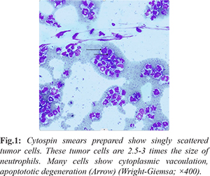
Contrast-enhanced computed tomography (CECT) of the thorax, neck, and paranasal sinuses demonstrated postoperative changes and reactive sinus changes, without evidence of pulmonary parenchymal abnormalities. Ultrasonography of the abdomen was normal. Nasal cavity examination revealed postoperative changes without evidence of tumor regrowth. Nerve conduction studies were within normal limits. Magnetic resonance imaging (MRI) of the spine revealed diffuse leptomeningeal enhancement along the dorsal and lumbar spinal cord, with thickening and enhancement of the dorsal nerve roots and cauda equina [Fig.2-4]. MRI of the brain was unremarkable. Based on the clinical presentation, CSF cytological findings, and spinal MRI features, a diagnosis of leptomeningeal carcinomatosis secondary to undifferentiated sinonasal carcinoma was established.
Discussion
Sinonasal undifferentiated carcinoma (SNUC) is a rare, highly aggressive malignancy characterized by rapid clinical progression and a propensity for central nervous system (CNS) invasion. However, leptomeningeal dissemination in SNUC is rarely reported compared with its occurrence in solid organ malignancies, melanoma, and hematological cancers [ 2, 3]. Tumor cells may reach the leptomeninges through several routes, including haematogenous dissemination via arterial circulation, extension from cerebral parenchymal or choroid plexus involvement, and direct spread from adjacent bony structures. Tumors arising in the palate and paranasal sinuses may also extend directly to the meninges, while perineural invasion and lymphatic dissemination represent additional potential pathways [ 4]. Leptomeningeal carcinomatosis can manifest with diverse neurological symptoms, including headache, altered mental status, nausea, vomiting, diplopia, cerebellar dysfunction, back pain, and lower limb weakness. Reported clinical manifestations include radicular pain (58%), altered mental status (49%), weakness (47%), headache (32%), neck or back pain (17%), and seizures (13%) [5,6]. The variability of these presentations can make early diagnosis challenging, particularly when neurological symptoms occur in the absence of detectable systemic disease. In the present case, the patient developed progressive paraparesis following treatment for SNUC with chemotherapy and radiotherapy. Notably, PET-CT performed 15 days after the onset of lower limb weakness showed no evidence of increased metabolic activity at the primary site or elsewhere. This finding highlights the possibility of leptomeningeal dissemination despite the absence of detectable metabolic activity on systemic imaging. A similar case was reported by Meng et al., in which a patient developed paraparesis one year after receiving radiotherapy and cisplatin-etoposide chemotherapy for SNUC [ 3]. Leptomeningeal carcinomatosis presenting as cauda equina syndrome has been more frequently described in association with melanoma, small-cell lung carcinoma, and lung adenocarcinoma [ 7- 10]. The present case highlights this uncommon neurological presentation in a patient with SNUC. In a retrospective study conducted in a single province, Xu et al. identified bone, cervical lymph nodes, lungs, brain, and liver as the most common metastatic sites of SNUC, in decreasing order of frequency [ 11]. The rarity of leptomeningeal involvement further emphasizes the importance of considering this diagnosis in patients with SNUC who develop new neurological deficits, even in the absence of evidence of primary tumor recurrence or other systemic metastases.
Conclusion
Sinonasal undifferentiated carcinoma (SNUC) is a rare and highly aggressive malignancy. Although leptomeningeal carcinomatosis secondary to SNUC is uncommon, it should be considered in the differential diagnosis of patients presenting with new neurological manifestations, including headache, cranial nerve deficits, or cauda equina syndrome, even in the absence of detectable primary tumor recurrence.
Contributors: AS, AH: found the concept and participated in writing; UK: revised and approved the final manuscript; JS: participated in writing. AS will act as a study guarantor. All authors approved the final version of this manuscript and are responsible for all aspects of this study. Funding: None; Competing interests: None stated.
References - Nguyen A, Nguyen A, Dada OT, Desai PD, Ricci JC, Godbole NB, et al. Leptomeningeal metastasis: A review of the pathophysiology, diagnostic methodology, and therapeutic landscape. Curr Oncol. 2023;30:5906-5931.
- Yust-Katz S, Cachia D, Kamiya-Matsuoka C, Theeler BJ, Olar A, Prado M, et al. Ependymomas arising outside of the Central Nervous System: A case series. Neuro-Oncol. 2016;18:vi162.
- Meng Y, Brunckhorst K, Filippi CG, Langer DJ, Goenka A, Teckie S. Spinal leptomeningeal metastasis of sinonasal undifferentiated carcinoma. Appl Radiat Oncol. 2018;7:44-47.
- Kokkoris CP. Leptomeningeal carcinomatosis: How does cancer reach the pia-arachnoid? Cancer. 1983;51:154-160.
- Kaplan JG, DeSouza TG, Farkash A, Shafran B, Pack D, Rehman F, et al. Leptomeningeal metastases: comparison of clinical features and laboratory data of solid tumors, lymphomas and leukemias. J Neurooncol. 1990;9:225-229.
- Clarke JL, Perez HR, Jacks LM, Panagaes KS, Deangelis LM. Leptomeningeal metastases in the MRI era. Neurology. 2010;74:1449-1454.
- Inci S, Bozkurt G, Gulsen S, Firat P, Ozgen T. Rare cause of subarachnoid hemorrhage: spinal meningeal carcinomatosis. Case report. J Neurosurg Spine. 2005;2:79-82.
- Noake JR, Shepherd A, Smith WR. Melanomatous leptomeningeal carcinomatosis masquerading as Guillain-Barré syndrome. Acute Med. 2010;9:20-23.
- Pagano A, Iaquinandi A, Fraioli MF, Bossone G, Carra N, Salvati M. Cauda equina syndrome from intradural metastasis of a non-neural tumor: case report and review of literature. Br J Neurosurg. 2023;37:1487-1494.
- Sun MG, Kim IY, Kim YJ, Jung TY, Moon KS, Jung S, et al. Lorlatinib therapy for rapid and dramatic control of brain and spinal leptomeningeal metastases from ALK-positive lung adenocarcinoma. Brain Tumor Res Treat. 2021;9:100-105.
- Xu CC, Dziegielewski PT, McGaw WT, Seikaly H. Sinonasal undifferentiated carcinoma (SNUC): the Alberta experience and literature review. J Otolaryngol - Head Neck Surg. 2013;42(1):2.
|