Introduction
Periampullary carcinomas arising from the ampulla of Vater, distal common bile duct, head of pancreas, or periampullary duodenum account for the majority of indications for Whipple’s pancreaticoduodenectomy. While surgical specimens from such procedures undergo meticulous pathological examination, incidental findings of significant diagnostic importance may occasionally be encountered. Accessory spleens (splenunculi) are common congenital anomalies, present in approximately 10-30% of the general population, most frequently at the splenic hilum. Intrapancreatic accessory spleens (IPAS) are far less prevalent, occurring in approximately 1-2% of individuals, predominantly in the tail of the pancreas [
1,
2]. An even rarer phenomenon is the occurrence of an epidermoid cyst within such an intrapancreatic accessory spleen-the entity termed ECIPAS (Epidermoid Cyst in Intrapancreatic Accessory Spleen). ECIPAS is a benign entity whose imaging characteristics may closely mimic pancreatic cystic neoplasms, including serous cystadenoma, mucinous cystic neoplasm, or intraductal papillary mucinous neoplasm, leading to potential overdiagnosis and unnecessary surgery [
3].
The co-existence of ECIPAS with a synchronous malignant neoplasm in the same pancreaticoduodenectomy specimen has, to the best of our knowledge, not been previously documented. We herein describe such a case and discuss its clinicopathological features, diagnostic pitfalls, and the role of CD8 immunohistochemistry in establishing the correct diagnosis.
Case Report
A 46-year-old male presented with a 4-month history of progressive abdominal pain, jaundice, and pruritus. There was no associated fever, weight loss, or gastrointestinal bleeding. His past surgical history was significant for laparoscopic cholecystectomy performed 2 years earlier for chronic cholecystitis. He also had a 10-year history of chronic alcohol consumption and cigarette smoking. Physical examination revealed icterus and mild epigastric tenderness. Biochemical investigations showed elevated serum bilirubin, predominantly direct, raised alkaline phosphatase, and mildly elevated transaminases. Ultrasonography and contrast-enhanced computed tomography (CECT) of the abdomen revealed a periampullary mass. Endoscopic biopsy demonstrated moderately differentiated adenocarcinoma consistent with periampullary carcinoma. Following multidisciplinary team review, the patient underwent classical Whipple’s pancreaticoduodenectomy.
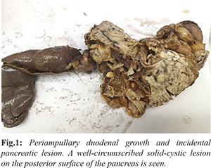
The pancreaticoduodenectomy specimen was received in its entirety. Gross examination revealed a greyish-white, firm, infiltrative growth involving the periampullary region of the duodenal mucosa. The cut surface was homogeneous with irregular margins. The common bile duct and main pancreatic duct appeared unremarkable, without intraluminal growth or dilatation. Regional lymph nodes were identified and submitted entirely for histopathological examination [Fig.1]. Incidentally, a separate, well-circumscribed solid-cystic lesion was identified on the posterior surface of the pancreas. The cyst contained clear serous fluid and had a smooth internal lining. The surrounding tissue showed a rim of reddish-brown parenchyma resembling splenic tissue.
Microscopic examination of the duodenal lesion revealed moderately differentiated adenocarcinoma composed of irregular glandular structures lined by pleomorphic epithelial cells with hyperchromatic nuclei, prominent nucleoli, and increased mitotic activity. Perineural invasion was absent, the surgical resection margins were free of tumour, and the regional lymph nodes were negative for metastatic adenocarcinoma. Sections from the pancreatic cystic lesion showed a cyst lined by stratified squamous epithelium. The cyst wall contained dense lymphoid tissue with well-formed follicles and reactive germinal centres. The interfollicular stroma exhibited sinusoidal spaces lined by flattened endothelial cells. No mucinous epithelium, papillary projections, ovarian-type stroma, or communication with the pancreatic ductal system was identified. The surrounding pancreatic parenchyma was unremarkable [Fig.2,3].
Immunohistochemical examination of the cystic lesion demonstrated CD8 positivity highlighting the sinusoidal spaces within the cyst wall, confirming the presence of splenic sinusoidal tissue characteristic of an intrapancreatic accessory spleen [Fig.4]. The squamous epithelial lining was positive for cytokeratin (CK) [Fig.5]. The combined histomorphological and immunohistochemical findings established the diagnosis of an epidermoid cyst in an intrapancreatic accessory spleen (ECIPAS), incidentally identified in a pancreaticoduodenectomy specimen containing periampullary adenocarcinoma.
Discussion
Accessory spleens are found in approximately 10-30% of individuals, most commonly at the splenic hilum, while intrapancreatic accessory spleens account for a small proportion (approximately 1-2%) and are predominantly located in the pancreatic tail [
1,
2]. Epidermoid cyst in intrapancreatic accessory spleen (ECIPAS), first described by Davidson et al. in 1980, is an exceptionally rare manifestation of this developmental anomaly. Fewer than 100 cases have been reported in the literature, with nearly all occurring in the pancreatic tail [
3,
4].
The pathogenesis of epidermoid cysts arising within accessory spleens remains unclear. The most widely accepted hypothesis suggests that mesothelial cell inclusions become entrapped within the developing splenic parenchyma during embryogenesis and subsequently undergo squamous metaplasia. Histologically, ECIPAS is characterised by a cyst lined by stratified squamous epithelium, surrounded by lymphoid tissue containing follicles and germinal centres, along with splenic sinusoidal spaces within the cyst wall. These features facilitate its distinction from other pancreatic cystic lesions.
The principal diagnostic challenge associated with ECIPAS is its resemblance to pancreatic cystic neoplasms, including mucinous cystic neoplasms, serous cystadenomas, solid pseudopapillary neoplasms, and cystic pancreatic neuroendocrine tumours [5]. On cross-sectional imaging, the peripheral solid component typically demonstrates enhancement similar to that of the normal spleen across different vascular phases. Endoscopic ultrasound-guided fine-needle aspiration (EUS-FNA) may yield serous fluid with low carcinoembryonic antigen (CEA) and amylase levels, while cytological examination generally lacks mucinous or neoplastic cells [6]. Radionuclide scintigraphy using technetium-99m sulphur colloid or heat-damaged red blood cells can identify functioning splenic tissue and aid diagnosis, although these techniques are infrequently employed [7]. Immunohistochemistry plays an important role in confirming the splenic origin of the lesion. CD8 immunostaining highlights the endothelial cells lining splenic sinusoids, providing a useful diagnostic marker for splenic tissue [8]. In the present case, CD8 positivity highlighted the sinusoidal spaces within the cyst wall, supporting the diagnosis of ECIPAS. This characteristic immunohistochemical pattern serves as a valuable adjunct to the morphological findings in establishing the diagnosis [8]. The present case is noteworthy because, to the best of our knowledge, it represents a rare occurrence of synchronous ECIPAS and periampullary adenocarcinoma within a single pancreaticoduodenectomy specimen. The periampullary adenocarcinoma was the primary indication for surgery, whereas ECIPAS was identified incidentally during histopathological examination. The absence of abnormalities in the common bile duct and main pancreatic duct further supported the periampullary origin of the carcinoma.
Recognising ECIPAS is clinically important because of its benign nature. Misinterpretation as a pancreatic cystic neoplasm may lead to unnecessary surgical intervention. In the present case, the lesion was incidentally resected during Whipple’s pancreaticoduodenectomy performed for periampullary adenocarcinoma and did not influence surgical management. Nevertheless, accurate histopathological characterisation is essential to establish the diagnosis, guide appropriate clinical counselling, and exclude an associated malignant process.
Conclusion
We report a documented case of synchronous periampullary adenocarcinoma and ECIPAS within a single Whipple’s pancreaticoduodenectomy specimen. Comprehensive histopathological evaluation of all lesions encountered in complex surgical specimens is imperative. ECIPAS should be included in the differential diagnosis of pancreatic cystic lesions, particularly those with a peripherally enhancing soft tissue component on imaging, to prevent unnecessary radical surgery. CD8 immunohistochemistry is a valuable and reliable ancillary tool for confirming the accessory splenic nature of such lesions.
Contributors: HS: Histopathological examination, immunohistochemical interpretation, manuscript drafting. AS: Literature review, manuscript drafting, critical revision. AM: Clinical management, critical revision. HS will act as a study guarantor. All authors have read and approved the final manuscript.
Funding: None; Competing interests: None stated.
References
- Horton KM, Hruban RH, Yeo CJ, Fishman EK. Multi-detector row CT of pancreatic islet cell tumors. Radiographics. 2006;26(2):453-464.
- Kimura W, Nagai H, Kuroda A, Muto T, Esaki Y. Analysis of small cystic lesions of the pancreas. Int J Pancreatol. 1995;18(3):197-206.
- Pal'yanov A, Borodach AV, Prokhorova IV. Epidermoid cyst in intrapancreatic accessory spleen. J Gastrointest Surg. 2014;18(7):1391-1393.
- Davidson ED, Campbell WG Jr, Hersh T. Epidermoid splenic cyst occurring in an intrapancreatic accessory spleen. Am J Gastroenterol. 1980;74(3):161-163.
- Valsangkar NP, Morales-Oyarvide V, Thayer SP, Ferrone CR, Wargo JA, Warshaw AL, et al. 851 resected cystic tumors of the pancreas: a 33-year experience at Massachusetts General Hospital. Surgery. 2012;152(3 suppl 1):S4-S12.
- Tan YM, Ooi LL, Mack PO. Epidermoid cyst arising from intrapancreatic accessory spleen: a case report and a review. Hepatogastroenterology. 2000;47(34):1167-1169.
- Holalkere NS, Hahn PF, Sahani DV. Intrapancreatic accessory spleen: diagnosis with echo-planar MRI. AJR Am J Roentgenol. 2006;186(2):347-351.
- Bruno C, Minniti S, Bucci A, Pozzi-Mucelli R. MDCT differentiation of hypervascular liver lesions in patients with cirrhosis: comparison of intrapancreatic accessory spleen with hypervascular liver cancer. AJR Am J Roentgenol. 2004;183(3):711-718.