|
|
|
|
|
Piperacillin–Tazobactam–Induced Type I Kounis Syndrome in a Young Male
|
|
|
|
Caroline Joseph Soans, Santosh Haridas Vhatkar, Amol Govind Chavhan G Plus Heart Hospital, Kalyan, Maharashtra 421301, India. |
|
|
|
|
|
Corresponding Author:
|
|
Dr Caroline Joseph Soans Email: carolsoans04@gmail.com |
|
|
|
|
|
|
|
|
Received:
16-OCT-2025 |
Accepted:
05-MAR-2026 |
Published Online:
25-JUN-2026 |
|
|
|
|
|
|
|
Abstract
|
|
|
|
Background: Kounis syndrome (KS), or allergic acute coronary syndrome, describes the concurrence of acute coronary events with hypersensitivity reactions. It remains underdiagnosed, particularly in patients without conventional cardiovascular risk factors. Case Report: We describe a 28-year-old male with no prior cardiac risk factors who developed severe anaphylaxis and acute left ventricular dysfunction immediately after receiving intravenous piperacillin-tazobactam (4.45g). Electrocardiogram showed biphasic T-wave changes, and echocardiography revealed severe global left ventricular systolic dysfunction (ejection fraction 15-20%). Emergency coronary angiography demonstrated normal epicardial coronary arteries. The combination of acute chest pain, ECG abnormalities, echocardiographic dysfunction, normal coronaries, and a temporal link to anaphylaxis confirmed the diagnosis of Type I KS. The patient improved with aggressive resuscitation, antihistamines, corticosteroids, cautious use of epinephrine, and supportive cardiac care. Conclusion: This case highlights the clinical significance of KS in the differential diagnosis of acute coronary syndromes precipitated by allergic reactions, especially in young individuals without cardiovascular risk factors. Early recognition and appropriate management are crucial, especially in young patients presenting with cardiac symptoms during anaphylaxis. |
|
|
|
|
|
Keywords :
|
Acute Coronary Syndrome; Anaphylaxis; Coronary Angiography; Echocardiography; Hypersensitivity.
|
|
|
|
|
|
|
|
|
|
|
|
Introduction
Kounis syndrome, first described by Nicholas Kounis and Zavras in 1991, refers to the concurrent occurrence of acute coronary syndrome and hypersensitivity reactions following an allergenic event [ 1]. Common triggers include antibiotics and nonsteroidal anti-inflammatory drugs (NSAIDs), as well as environmental exposures and insect stings [ 2]. The three variants of the condition are: Type I refers to coronary artery spasm in patients with angiographically normal coronary arteries; Type II involves acute plaque rupture or erosion in those with pre-existing atherosclerosis; and Type III refers to coronary stent thrombosis or restenosis precipitated by allergic reactions [ 3]. Among these variants, Type I is the rarest but potentially life-threatening condition and is often ignored in young patients with no cardiovascular risk factors. Studies have reported that KS is underdiagnosed, despite its true prevalence being unknown. Among those experiencing allergic reactions, between 1.1% and 3.4% of people reported symptoms; however, many cases may go unnoticed due to low awareness and nonspecific cardiac signs [1]. Chest pain occurring during anaphylaxis is often attributed only to hemodynamic instability, which can delay recognition of concurrent coronary involvement. This highlights the attention to younger patients to consider KS without conventional cardiovascular risk factors, as its symptoms can resemble those of other acute coronary diseases, which could result in misdiagnosis or delayed treatment [4]. Management requires simultaneous treatment of both the allergic reaction and the acute coronary event. This approach is usually complex. Eliminating the offending allergen and initiating corticosteroids, antihistamines, and intravenous fluids are essential. The first-line treatment for anaphylaxis is epinephrine; however, its use is restricted because it can worsen coronary vasospasm by stimulating the a-adrenergic receptor. Titrated doses or intramuscular injection are recommended over high-dose intravenous infusion in certain situations [5]. From the cardiac perspective, vasodilators such as nitrates and calcium channel blockers are effective for relieving coronary vasospasm [6]. In contrast, ß-blockers are avoided because they worsen anaphylaxis by inhibiting ß-adrenergic pathways essential for bronchodilation [7]. Multidisciplinary management involving cardiologists, emergency physicians, and allergists is recommended to tailor treatment and prevent recurrence. Documenting the allergen, providing patient counselling, and setting up a referral for allergy testing or desensitisation are advised. However, there are no strong standardised guidelines for treatment; hence, decisions often rely on case reports and clinician experience, resulting in variability and potential risks [3]. We present an uncommon case of piperacillin-tazobactam-induced Type I KS leading to severe systemic anaphylaxis with acute left ventricular failure in a young healthy male. The unique feature was the rapid onset of fulminant anaphylaxis within a few minutes of drug infusion, associated with severe global left ventricular systolic dysfunction to an ejection fraction of 15-20%. Within four days, echocardiography demonstrated partial recovery, with the ejection fraction improving to 40%. Such severe ventricular dysfunction with angiographically normal coronaries is extremely uncommon and has important implications.
Case Report
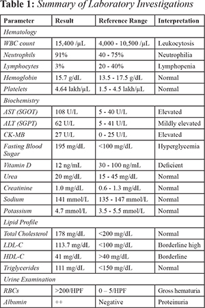
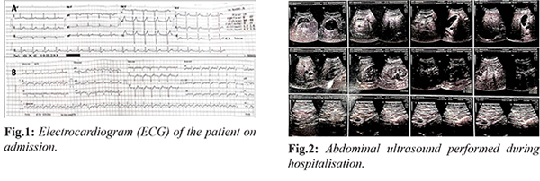
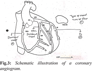
A 28-year-old previously healthy male with no family history of cardiovascular or allergic disorders underwent double-J (DJ) stent exchange with retrograde intrarenal surgery (RIRS) for renal calculi. Post-procedure, he developed a fever and was started on ceftriaxone. Due to persistent fever, antibiotics were escalated to intravenous piperacillin-tazobactam. Within minutes of drug administration (4.45g), he experienced severe anaphylaxis with hypotension (systolic BP <80 mmHg), generalised erythematous rash, and respiratory distress and acute retrosternal chest pain with diaphoresis. Electrocardiography revealed sinus tachycardia with bifid T-waves in V1 and V2. Two-dimensional echocardiography revealed severe global hypokinesia of the LV, which was predominant in the anterior, anterolateral, and anteroseptal walls at an estimated LVEF of 15-20%. Cardiac biomarkers revealed a minimally elevated troponin I. Coronary angiography was performed urgently to rule out acute coronary syndrome, revealing angiographically normal coronary arteries with no obstructive lesions or atherosclerosis. The concurrence of an acute allergic reaction, ECG changes, reversible left ventricular dysfunction, and normal coronary arteries confirmed Type I Kounis syndrome. The patient was evaluated through laboratory biomarkers, imaging, and cardiac function tests to establish the diagnosis and assess organ function. All reports are summarised in Table 1. Initial laboratory results showed leukocytosis with a total WBC of 15,400/µL and marked neutrophilia (91%), along with relative lymphopenia (3%), indicating an acute systemic inflammatory and allergic response. Hemoglobin (15.7 g/dL), platelet count (464,000/µL), and red cell indices were normal. Follow-up counts normalised, though mild eosinophilia persisted, further supporting an allergic cause. Cardiac enzymes revealed a slight increase in CK-MB (27 U/L) and a minimally elevated troponin I level, suggestive of myocardial infarction. Biochemistry demonstrated a mild rise in transaminases (AST 108 U/L, ALT 62 U/L), with normalised bilirubin and protein levels, consistent with a short hepatocellular stress likely secondary to anaphylaxis. Renal function was unchanged. The lipid profile showed total cholesterol at 178 mg/dL, LDL at 113.7 mg/dL, triglycerides at 111 mg/dL, and HDL at 41 mg/dL, which suggests borderline dyslipidemia. Fasting blood sugar was 195 mg/dL, consistent with diabetes. Vitamin D was low at 12 ng/mL, pointing to further metabolic issues. Urine examination demonstrated gross haematuria (>200 RBCs/HPF) and proteinuria (++ albumin), findings attributed to the patient’s underlying renal calculi and recent stent exchange rather than the acute cardiac event. Serial echocardiograms gave useful diagnostic details. The initial test showed severe global hypokinesia of the left ventricle, with an ejection fraction between 15 and 20%. On follow-up after four days, the repeat echocardiogram showed partial improvement, with ejection fraction rising to 40 to 45%. The heart valves appeared normal, with no signs of diastolic dysfunction, pulmonary hypertension, or pericardial effusion. Electrocardiographic changes were consistent with acute coronary involvement. Fig.1a represents the ECG at presentation with marked ST-segment elevation in anterior precordial leads, consistent with acute myocardial injury during anaphylaxis. A subsequent ECG [Fig.1b] demonstrated evolving changes with attenuation of ST-segment elevation, in line with clinical recovery. Fig.2 represents the abdominal ultrasound performed during hospitalisation. A coronary angiogram was performed to exclude structural coronary disease. Fig.3 represents the angiogram revealed completely normal epicardial coronaries with TIMI-III flow, confirming the diagnosis of Type I KS (vasospastic mechanism without atherosclerotic obstruction).
The patient was promptly transferred to the intensive cardiac care unit. Hypotension was managed with intravenous fluids. Intravenous antihistamines and corticosteroids were administered. Caution was taken when administering epinephrine because it could have exacerbated coronary vasospasm. The causative agent, piperacillin-tazobactam, was immediately discontinued. Immediately, oxygen was administered along with continuous monitoring. Calcium channel blockers and nitrates were used to relieve coronary spasm. Beta-blockers were avoided due to the risk associated with anaphylaxis. The recovery of left ventricular (LV) function was monitored by serial echocardiography. The patient’s condition stabilised over time, with progressive improvement in hemodynamics and symptoms. Follow-up echocardiography after four days indicated partial restoration of left ventricular function, with ejection fraction improving to 40%.
Discussion
KS was described as an allergic angina, which is a rare but fatal condition due to the co-incidental occurrence of an acute coronary syndrome with hypersensitivity reactions [ 8]. The management of the syndrome depends on the types, such as Type I, which usually requires conservative management with antiallergic and vasodilator therapy, whereas Types II and III often necessitate standard acute coronary syndrome protocols, including antiplatelets, anticoagulation, and, in some cases, revascularisation [ 2, 9]. The first immediate action is discontinuation of the offending allergen, followed by corticosteroids, antihistamines, and intravenous fluids. Although epinephrine remains the first-line therapy, it is advised to use it cautiously and preferably intramuscularly, as intravenous administration may aggravate coronary vasospasm [ 9, 10]. Our case aligns with previous reports which has been associated with antibiotic-induced KS [4,11,12]. Evidence reported that antibiotics such as amoxicillin-clavulanate and ciprofloxacin acted as triggers, with a rapid onset of allergic symptoms followed by ischemic ECG changes, followed by resolution with anti-allergic and vasodilator/supportive therapy. However, what distinguishes our case is the extreme severity of myocardial dysfunction (LVEF ~ 15-20%), its immediate onset after exposure to the antibiotic, the intensity of systemic anaphylaxis, and the documented partial recovery to ~ 40% LVEF in a young male. Most cases show normal coronaries and reversible outcomes, confirming vasospasm as the primary mechanism. The management of KS is challenging as physicians have to simultaneously treat allergic reaction as well as an acute coronary event. Nayoshi et al. [4] reported management of a young woman with Type I KS by intramuscular epinephrine 0.3 mg, intravenous corticosteroids, and antihistamines for anaphylaxis. Also, for coronary vasospasm, sublingual nitroglycerine was administered, leading to complete recovery. Similarly, Chatterjee et al. [13] demonstrated successful treatment of a middle-aged woman with amoxicillin–clavulanate–induced KS using intravenous hydrocortisone (100 mg), intravenous pheniramine (45 mg), and intramuscular epinephrine (0.5 mg). In another study by Tang et al. [14], patient developed limb erythema, hypotension, and chest tightness after receiving intravenous flurbiprofen. She was successfully managed with intravenous methylprednisolone 80 mg, diphenhydramine 50 mg, and isosorbide dinitrate 5 mg sublingual. From a cardiac point of view, these reports consistently revealed that vasodilators such as nitrates and calcium channel blockers are effective for relieving coronary vasospasm, whereas ß-blockers should be avoided because of their potential to aggravate anaphylaxis. In this present case, epinephrine was cautiously administered along with corticosteroids, antihistamines, and intravenous fluids, while ß-blockers were avoided. This case highlights the importance of early detection, accurate diagnosis, and individualised therapy in Kounis syndrome. Prompt withdrawal of the allergen, careful balance of anaphylaxis and cardiac management, and multidisciplinary collaboration are essential for favourable outcomes.
Conclusion
This case highlights the necessity of considering Type I KS as a cause of acute cardiac involvement in anaphylaxis, even in patients with no cardiac risk factors and angiographically normal coronaries. Early recognition can prevent misdiagnosis and inadequate management, needing a careful consideration of life-saving anaphylaxis therapy compared with interventions to mitigate the effects of coronary vasospasm. Physician awareness is necessary for the successful treatment of this rare but potentially fatal condition.
Contributors: CJS was responsible for conceptualisation, methodology, and manuscript writing. SHV and AGC contributed to drafting of the original manuscript. Data curation, investigation, methodology, and visualisation were performed by all authors. CJS will act as a study guarantor. All authors participated in the review and editing of the manuscript and approved the final version for publication. Funding: None; Competing interests: None stated.
References - Renda F, Landoni G, Trotta F, Piras D, Finco G, Felicetti P, et al. Kounis Syndrome: An analysis of spontaneous reports from international pharmacovigilance database. Int J Cardiol. 2016;203:217-220.
- Abdelghany M, Subedi R, Shah S, Kozman H. Kounis syndrome: A review article on epidemiology, diagnostic findings, management and complications of allergic acute coronary syndrome. Int J Cardiol. 2017;232:1-4.
- Fassio F, Losappio L, Antolin-Amerigo D, Peveri S, Pala G, Preziosi D, et al. Kounis syndrome: A concise review with focus on management. Eur J Intern Med. 2016;30:7-10.
- Nanyoshi M, Hayashi T, Sugimoto R, Nishisaki H, Kenzaka T. Type I Kounis syndrome in a young woman without chest pain: a case report. BMC Cardiovasc Disord. 2024;24(1):467.
- Omri M, Kraiem H, Mejri O, Naija M, Chebili N. Management of Kounis syndrome: two case reports. J Med Case Reports. 2017;11(1):145.
- Fourie P. Kounis syndrome: a narrative review. South Afr J Anaesth Analg. 2016;22(2):72-80.
- Fang W, Song L, Deng Z, Sun W, Li Z, Wang C. Analysis of clinical features of Kounis syndrome induced by Cephalosporin. Front Cardiovasc Med. 2022;9:885438.
- Kounis syndrome: A manifestation of drug-eluting stent thrombosis associated with allergic reaction to contrast material - ScienceDirect. Available from: https://www.sciencedirect.com/science/article/abs/pii/S0167527308009856. Accessed on October 16, 2025
- Fassio F, Losappio L, Antolin-Amerigo D, Peveri S, Pala G, Preziosi D, et al. Kounis syndrome: A concise review with focus on management. Eur J Intern Med. 2016;30:7-10.
- Kounis NG. Kounis syndrome (allergic angina and allergic myocardial infarction): a natural paradigm? Int J Cardiol. 2006;110(1):7-14.
- Srouji SM, Haroun Y, Vemuri S, Bakhsh MU. Kounis Syndrome triggered by medications and an illicit substance: A report of a rare Ccse. Cureus. 2025; Available from: https://www.cureus.com/articles/348606-kounis-syndrome-triggered-by-medications-and-an-illicit-substance-a-report-of-a-rare-case. Accessed on October 16, 2025
- Shiza S. Type 1 Kounis Syndrome: Allergic vasospastic cardiac event triggered by Ciprofloxacin. Am J Prev Cardiol. 2024;19:100765.
- Chatterjee S, Paranjape S, Singh G. Kounis Syndrome after Oral Amoxicillin Clavulanate. Int J Appl Basic Med Res. 2025;15(1):66-68.
- Tang C, Chen Y, Gu X. Case Report: Flurbiprofen-induced Type I Kounis syndrome. Front Cardiovasc Med. 2023;10:1284408.
|
|
|
|
|
|
|
Search Google Scholar for
|
|
|
Article Statistics |
|
Soans CJ, Vhatkar SH, Chavhan AGPiperacillin–Tazobactam–Induced Type I Kounis Syndrome in a Young Male.JCR 2026;16:50-54 |
|
Soans CJ, Vhatkar SH, Chavhan AGPiperacillin–Tazobactam–Induced Type I Kounis Syndrome in a Young Male.JCR [serial online] 2026[cited 2026 Oct 11];16:50-54. Available from: https://www.casereports.in/articles/16/2/Piperacillin-Tazobactam-Induced-Type-I-Kounis-Syndrome-in-a-Young-Male.html |

|
|
|
|
|